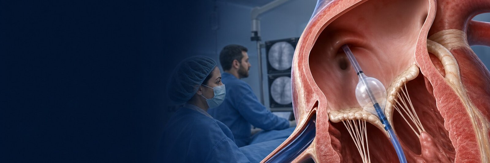
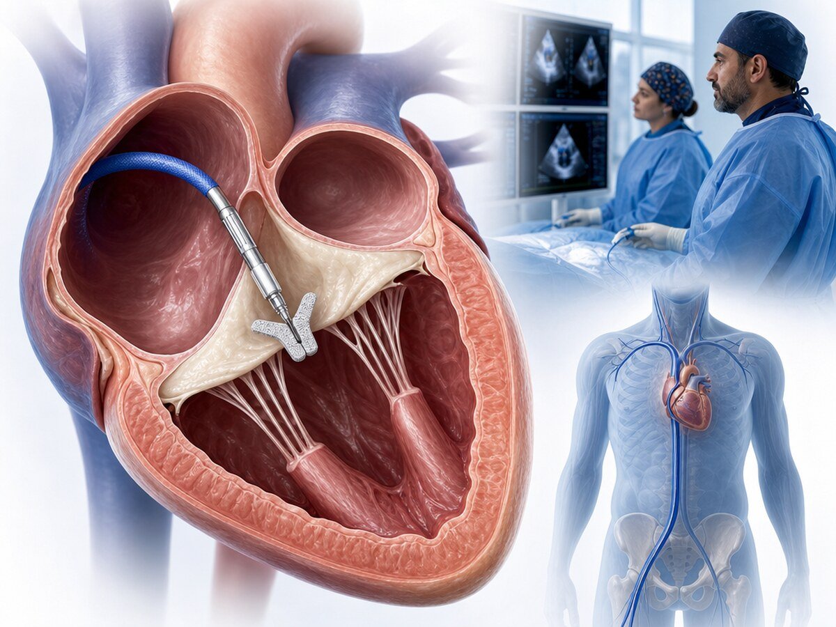
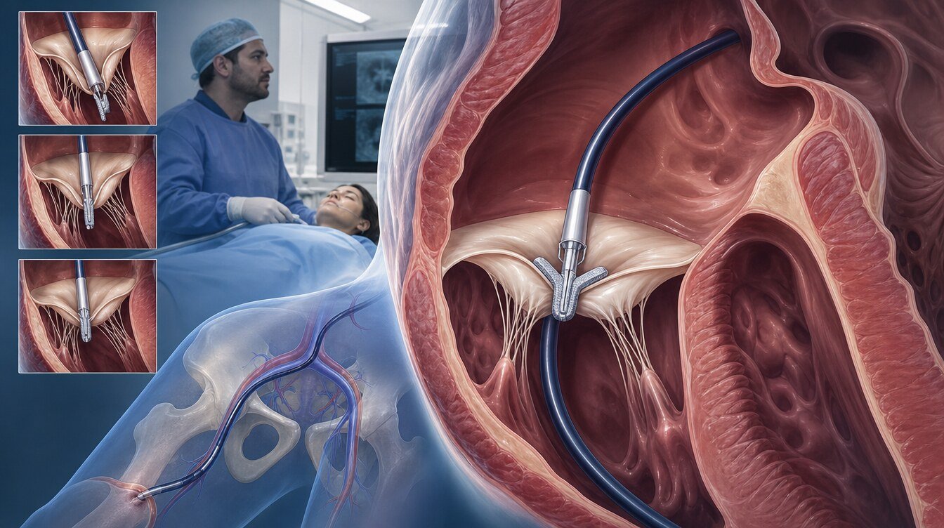
TriClip is a device system used for tricuspid transcatheter edge-to-edge repair (T-TEER) in selected patients with severe tricuspid regurgitation. The native valve is not removed; suitable leaflets are approximated from a femoral venous approach.[1–4]
What is tricuspid regurgitation?
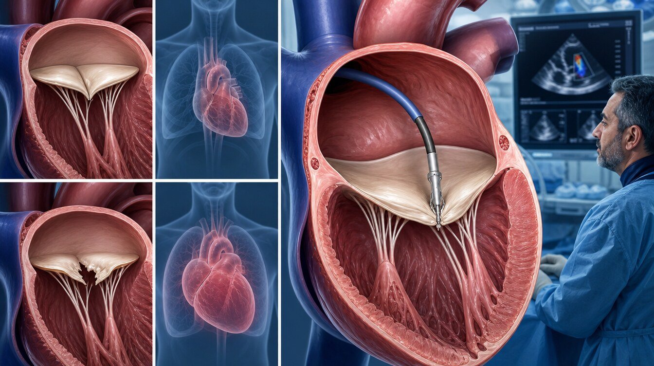
The tricuspid valve lies between the right atrium and ventricle. Regurgitation can cause oedema, abdominal congestion, fatigue and breathlessness. Mechanisms include primary leaflet disease, annular/right-heart enlargement, pulmonary hypertension and cardiac-device leads.[1,5]
Why does timing matter?
Severe TR can be followed by progressive right-ventricular dysfunction, venous congestion, liver or kidney involvement and frailty. If assessment occurs after advanced organ injury, reducing regurgitation may not translate into meaningful recovery. Early assessment means discussing options while recovery remains plausible; it does not mean intervening in everyone.[1]
Who may be assessed for TriClip?
The 2025 ESC/EACTS guideline supports transcatheter treatment in symptomatic isolated severe TR at increased surgical risk when anatomy is suitable, in the absence of severe RV dysfunction or precapillary pulmonary hypertension, to improve quality of life and right-heart remodelling.[1]
Important imaging questions include the mechanism and jet location, coaptation gap, leaflet length and motion, number of jets, baseline tricuspid gradient, pacing-lead interaction, right-ventricular function and pulmonary haemodynamics. No single measurement is a universal suitability threshold; three-dimensional imaging and, when needed, invasive haemodynamics are interpreted together.
What are the alternatives?
Medical therapy addresses congestion and underlying rhythm, left-heart or pulmonary disease but does not necessarily correct the anatomical gap. Surgery may be preferred when another cardiac operation is needed or anatomy favours repair/replacement. Other transcatheter repair or replacement strategies are separate procedures with different anatomical requirements. In advanced disease with little expected recovery, symptom-focused care may offer greater net benefit.[1]
How is TriClip performed?
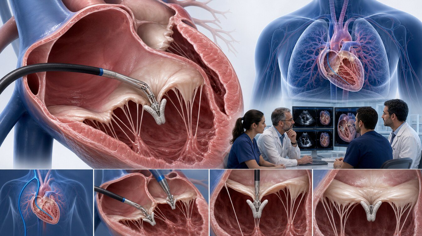
Venous access is obtained through the groin. The delivery system is advanced into the right atrium and aligned with the tricuspid valve using echocardiography and fluoroscopy. Target leaflets are captured, regurgitation and gradient are reassessed, and additional implants may be used. Unlike mitral TEER, no transseptal passage is required.
What does the evidence show?
TRILUMINATE Pivotal favoured T-TEER for a one-year hierarchical composite, driven mainly by quality-of-life improvement. The between-group KCCQ difference was 9.4 points at one month and 10.4 points at one year; death, tricuspid surgery and heart-failure hospitalisation did not appear different at one year.[2,6] Two-year results require interpretation in light of crossover.[3]
Tri.Fr randomised 300 anatomically suitable patients. At one year, the composite clinical and patient-reported outcome favoured T-TEER, with a 14.5-point between-group KCCQ difference. Its open-label design and selected anatomy limit generalisability.[4]
Is technical success the same as patient benefit?
No. Device implantation and lower TR are procedural goals. Clinical success also considers oedema, breathlessness, daily function, hospitalisation, organ congestion and quality of life. Residual TR, the capacity of the right ventricle to recover and comorbid disease influence the result. Trial averages are not individual promises.
Risks and follow-up
Risks include bleeding, vascular injury, cardiac injury, leaflet damage or single-leaflet attachment, residual/recurrent TR, tricuspid stenosis, device migration, rhythm disturbance, kidney injury, infection, stroke, reintervention and death. Pacing-lead interaction requires separate planning.[1,5]
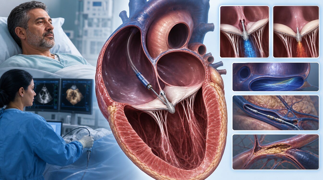
Recovery and medicine plans are individual. Diuretics are not automatically stopped. Oedema, weight, breathlessness, kidney function and echocardiographic findings remain under follow-up.
Long-term assessment includes residual or recurrent TR, tricuspid gradient, right-heart function, rhythm, organ function and whether clinical improvement is sustained.
Questions to ask the Heart Team
- What is causing the regurgitation, and does a pacing lead contribute?
- Is right-heart or organ injury still likely to recover?
- Does pulmonary vascular disease limit expected benefit?
- Which anatomical feature makes T-TEER favourable or difficult?
- What level of TR reduction and clinical improvement is the goal?
- Why is surgery or another transcatheter strategy not preferred?
- What is the next option if the result is incomplete?
Frequently asked questions
No. It is a catheter-based tricuspid T-TEER procedure, but it remains a major cardiac intervention.
They use a related edge-to-edge principle on different valves: TriClip targets the tricuspid valve and MitraClip the mitral valve.
Advanced right-heart failure, precapillary pulmonary hypertension, irreversible organ damage or unsuitable anatomy may limit benefit.[1]
Sometimes. The lead mechanism and relationship with the leaflets require detailed imaging.
Not automatically. Fluid status, kidney function and symptoms guide the individual plan.
Yes. Disease progression or leaflet/device changes can increase regurgitation; follow-up is required.
Timing depends on access-site findings, right-heart status and overall condition; follow the individual discharge plan.
Assessment and decision
The decision considers the chance of right-heart and organ recovery, anatomy, surgical alternatives and patient goals.
Literature
References
Praz F, et al. 2025 ESC/EACTS Guidelines. doi:DOI: 10.1093/eurheartj/ehaf194.
Sorajja P, et al. TRILUMINATE Pivotal. N Engl J Med. 2023. doi:DOI: 10.1056/NEJMoa2300525.
Kar S, et al. TRILUMINATE two-year outcomes. Circulation. 2025. doi:DOI: 10.1161/CIRCULATIONAHA.125.074536.
Donal E, et al. Tri.Fr. JAMA. 2025. doi:DOI: 10.1001/jama.2024.21189.
Hahn RT, et al. TVARC definitions. Eur Heart J. 2023. doi:DOI: 10.1093/eurheartj/ehad653.
Arnold SV, et al. Health Status After Transcatheter Tricuspid-Valve Repair. J Am Coll Cardiol. 2024. PMID 37898329.
This content is for general information only and does not replace personalized medical advice. In emergencies call local emergency services.
Continue