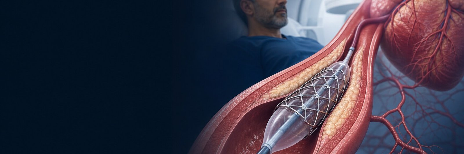
Peripheral artery disease (PAD) is a circulation disorder that most often appears when atherosclerosis narrows or blocks the arteries that carry blood to the legs. The disease may cause no symptoms; it may cause pain in the calf, thigh or buttock on walking; and in more advanced stages it may cause rest pain, a wound that will not heal or gangrene. PAD is not only a leg problem; it is also linked to the risk of heart attack and stroke.[1,2]
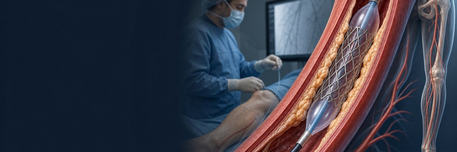
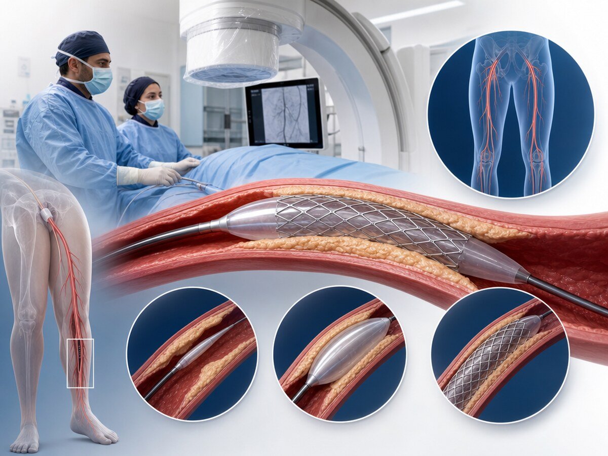
Stenting is the placement of a metal or coated scaffold to support the vessel wall from inside after a narrowed artery has been widened with a balloon. Not every person with PAD needs a stent. The decision depends on how severe the symptoms are, whether there is a wound or ischaemia, which arterial segment is involved, the vessel anatomy, accompanying diseases and the person’s goals.[1,2]
What is PAD and why does it matter?
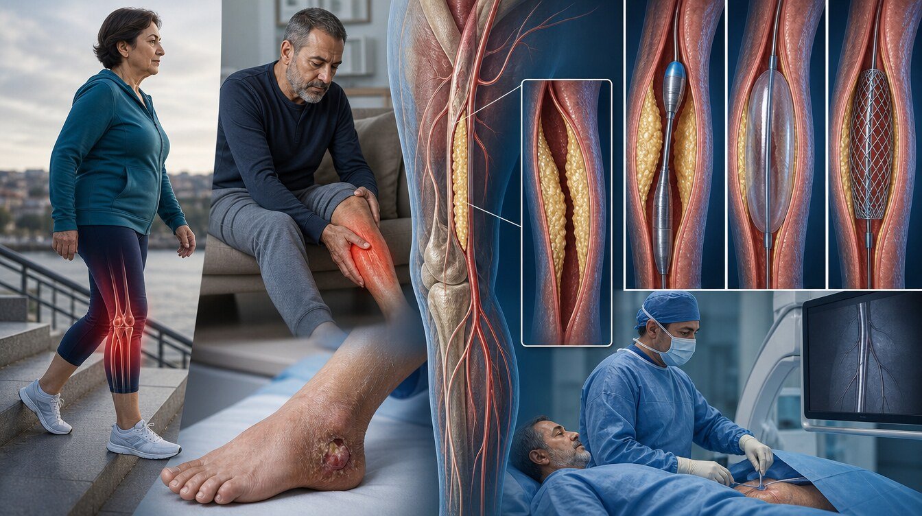
Peripheral artery disease is narrowing or blockage of arteries outside the heart, especially those of the legs. The most common cause is an atherosclerotic plaque of cholesterol, calcium, connective tissue and inflammatory cells in the vessel wall. As the inner diameter falls, the blood flow needed by working muscle during walking can no longer be met.
The leg arteries are grouped into different segments:
- Aortoiliac: the abdominal aorta and the iliac arteries around the pelvis.
- Femoropopliteal: the femoral artery that begins in the groin and the popliteal artery behind the knee.
- Infrapopliteal: the tibial and peroneal arteries from below the knee to the foot.
Calibre, bending load, how widespread the plaque is and which treatments can be used differ. An aortoiliac approach is not automatically transferred to femoropopliteal or below-knee disease.[1,2,12]
A PAD diagnosis also means the heart and brain arteries should be considered. The aim of care is not only to reduce leg pain or keep a vessel open; reducing the risk of heart attack, stroke, acute limb ischaemia and limb loss is part of the plan.[1,2]
Smoking, diabetes, high blood pressure, high cholesterol, kidney disease, older age, inactivity and previous cardiovascular disease are important risk factors. In diabetes and kidney failure, calcification can mislead diagnostic tests.[1,2]
PAD is not one picture. Asymptomatic PAD, claudication, chronic limb-threatening ischaemia (CLTI) and acute limb ischaemia are separate syndromes; their urgency and goals differ.[1,2]
This page is not the same disease as carotid artery stenosis. Carotid evidence and device results are not transferred to leg PAD.
Typical and atypical symptoms
The best-known symptom is claudication: muscle pain that starts with walking and eases with rest. The pain is usually felt in the muscle group below the narrowing. Hip or thigh pain may suggest aortoiliac disease; calf pain may suggest femoropopliteal disease. Below-knee disease can cause more widespread foot or calf symptoms.[1]
Typical claudication starts after a given distance, eases within a few minutes of stopping and returns when walking starts again. In more advanced disease there may be coldness, paleness or bluish colour in the foot, weak or absent pulses, a scrape that will not heal, a wound, blackening and rest pain that is worse at night.
Many people do not describe typical walking pain. Early tiring, weakness, a quietly shortening walking distance, pain only on hills, or a foot wound that will not heal without clear pain can be atypical. Hip, knee or back disease, nerve-root compression, neuropathy and venous disease can cause similar symptoms. The diagnosis does not rest on the pain description alone.[1,2]
Asymptomatic PAD
Asymptomatic PAD means arterial narrowing or a low arterial pressure is present without a clear leg complaint. The person may not walk far enough, or may have quietly limited activity. The diagnosis is often made during a pulse check, ultrasound or ABI.
The aim is not an automatic stent. The priority is to reduce cardiovascular risk, stop smoking, control cholesterol and blood pressure, manage diabetes, stay active and care for the feet. Routine revascularisation for a narrowing seen only on imaging is usually not appropriate.[1,2] A wound, necrosis, rest pain or rapid worsening means the picture is no longer asymptomatic.
Claudication
Claudication occurs when the extra oxygen demand of exercise cannot be met because the arteries are narrowed. Pain that eases with rest can help separate it from joint or nerve pain; it is not enough for a diagnosis on its own.
In most people the first step is comprehensive medical therapy and structured exercise. Exercise does not remove the plaque; it can improve walking tolerance and daily function.[1,7] If smoking continues, the benefit of exercise and medicine may fall.
Revascularisation may be considered when symptoms still clearly limit daily life despite lifestyle change, risk-factor control and exercise given for an adequate time. A narrowing on imaging is not, by itself, a reason for a stent.[1,2]
Chronic limb-threatening ischaemia (CLTI)
CLTI is an advanced picture in which a foot wound that has not healed for more than two weeks, gangrene or ischaemic rest pain is present together with objective circulatory impairment.[1,2] Rest pain is usually felt in the foot or toes and may worsen when the leg is horizontal. Diabetic neuropathy, infection and pressure sores can mask the picture.
CLTI needs prompt joint assessment by a vascular specialist and, when needed, diabetes, infection and wound-care teams. WIfI staging brings wound, ischaemia and foot infection together as a clinical frame for amputation risk and wound healing; staging is not, by itself, a treatment claim.[13]
The aim, in suitable people, is enough blood flow for tissue to heal, less pain and a functional limb. Technical vessel patency, wound healing, amputation-free survival and length of life are not the same outcomes.[1,3,4,13]
Acute limb ischaemia
Acute limb ischaemia is a sudden fall or stop in blood flow to the leg or foot. A clot, sudden occlusion, an embolus from the heart or loss of flow inside a stent or bypass can be among the causes.
Sudden severe pain, paleness, loss of pulses, coldness, numbness and weakness mean the person should not wait and should go to an emergency department. This is not a planned claudication complaint; assessment may be needed within hours.[1,2]
Diagnosis: ABI, exercise ABI, TBI, duplex, CTA/MRA and angiography
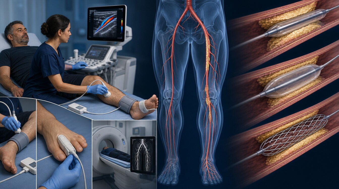
The ankle–brachial index (ABI) is the ratio of ankle systolic pressure to arm pressure. A low ABI supports meaningful flow limitation. In diabetes, chronic kidney disease and older age the vessel wall may become incompressible; the ABI can then be high or falsely normal. If clinical suspicion is high, a normal ABI does not rule the disease out.[1,2]
When the resting ABI is normal or borderline and there are walking symptoms, an exercise ABI can help. The toe–brachial index (TBI) and toe pressure are less affected by calcification than the ABI and can add to the assessment of tissue perfusion when CLTI is suspected.[1,2]
Duplex ultrasound shows flow and the site of narrowing without radiation. CTA gives detailed anatomy; kidney function and allergy history are reviewed because of iodinated contrast and radiation. MRA sometimes avoids radiation; implant and contrast limits apply. Catheter angiography is often chosen not only for diagnosis but when treatment in the same session is planned; it carries risks of bleeding, contrast reaction, kidney injury and vessel damage.[1,2]
Medical therapy and stopping smoking
Medical therapy continues whether or not a stent is placed: stopping tobacco, cholesterol and blood-pressure control, diabetes care, physical activity, antithrombotic therapy in suitable people, and foot and wound care.[1,2] Stopping smoking is a core part of PAD care. Cholesterol (hyperlipidaemia) treatment aims to protect the heart and brain arteries as well as the leg arteries. Medicines should not be started or stopped on a person’s own decision.
Structured exercise
In claudication, structured and, where possible, supervised exercise is a core component. The person walks under supervision, rests when symptoms appear and walks again when it is appropriate. A Cochrane review found that supervised exercise can increase walking distance compared with home-based exercise or walking advice alone.[7] Exercise does not remove plaque and does not replace medical therapy.
If there is heart disease, marked breathlessness, a balance problem or an open wound, a medical review is needed before a programme. Standard claudication exercise is not used in CLTI when there is an open wound or clear tissue loss.[1,7]
When is revascularisation considered?
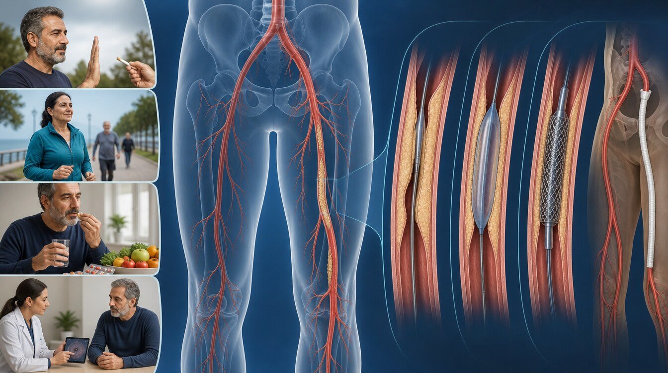
Endovascular treatment is done from inside the artery with a balloon, stent or another device; bypass creates a new flow path around the narrowed segment.
In claudication a procedure is considered when daily life remains unacceptably limited despite medical therapy and structured exercise.[1,2] In CLTI, if there is a wound, gangrene or ischaemic rest pain, revascularisation is often discussed without delay. In acute ischaemia the assessment is urgent.[1,2]
Angioplasty, stents and other devices
Angioplasty is widening of a narrowed segment with a balloon. A balloon alone is sometimes enough; a stent may be needed because the wall tends to recoil or because of a dissection. Bare-metal/nitinol stents, drug-eluting stents, drug-coated balloons, covered stent-grafts and atherectomy are different classes; they are not interchangeable.[1,2,12]
Femoropopliteal arteries carry a special mechanical load from knee motion; below the knee, small calibre and long disease stand out. A femoropopliteal device study does not automatically create a preference for aortoiliac or infrapopliteal disease. REAL PTX compared a drug-eluting stent with a drug-coated balloon in the femoropopliteal artery; it is not generalised to other segments.[12] Atherectomy is not routine and is not a universal step shown to improve hard clinical outcomes in all lesions.[1,2]
A mortality signal with drug-coated (usually paclitaxel) devices was opened in 2018; that historical concern is not closed by saying it “never existed”. The 2020 SWEDEPAD interim analysis found no difference in all-cause death.[8] A 2023 patient-level meta-analysis did not report an association between femoropopliteal drug-coated devices and death.[9] SWEDEPAD 1 (CLTI, 2025) did not reduce major ipsilateral amputation and showed no difference in death.[10] SWEDEPAD 2 (claudication, 2025) did not improve one-year quality of life; death was not significantly different over the whole follow-up, while five-year mortality incidence was higher in the coated-device arm.[11] A drug-coated device is therefore not the preferred option in every lesion; the choice follows current evidence, regulatory information and individual anatomy.
Evidence for a coronary drug-coated balloon or coronary stent is not transferred to the leg arteries.
Endovascular treatment or bypass?
The decision reviews the arterial segment and anatomical complexity, a target vessel to the foot, calcification, whether a suitable vein is available, heart, lung and kidney disease, frailty and the person’s goal (walking, wound healing, limb salvage).[1–4]
BEST-CLI showed that in CLTI, in the cohort with an adequate great saphenous vein (cohort 1), surgery improved the composite of a major limb event or death; in the cohort without an adequate vein (cohort 2) there was no significant difference between surgery and an endovascular approach.[3] BASIL-2 reported that, in CLTI needing infra-popliteal (± more proximal) revascularisation, a best-endovascular-first strategy had better amputation-free survival; the difference was driven largely by deaths.[4] These two trials are not the same population and do not mean that one method is superior in everyone.
How does the procedure proceed?
The frame below is not an operator manual.
- Symptoms, pulses, wounds, kidney function, bleeding risk, medicines and imaging are reviewed.
- The goal may be walking capacity, rest pain, wound healing or acute loss of flow.
- Local anaesthesia and, if needed, sedation are usually used; the catheter is advanced from the groin or another suitable artery.
- Contrast shows the site and length of the narrowing.
- A balloon, stent or another suitable device is used over a guidewire.
- Flow, vessel injury and clot are checked; the access site is closed and watched.
Contrast allergy, possible pregnancy, kidney disease and blood thinners should be told to the team. Stopping a medicine is only the treating team’s decision. Coronary angiography is a separate diagnostic and treatment path; it does not replace leg angiography.
Risks
Possible complications include access-site bleeding or haematoma, vessel rupture, sudden closure, embolisation, a contrast reaction, a fall in kidney function, clot inside a stent, restenosis, infection, rarely emergency surgery, heart attack, stroke and death.[1,2] Bypass can involve wound problems, graft occlusion and a longer recovery. Patency is not the same as wound healing, limb salvage or amputation-free survival.[1,3,4,13]
Antithrombotic therapy after the procedure
The aim is to reduce clot inside a stent or graft and cardiovascular events. In many people a single long-term antiplatelet medicine is the base approach; a short course of two antiplatelet medicines or another strategy may be chosen according to the procedure, other heart disease and bleeding risk.[1,2]
In selected suitable people, aspirin plus rivaroxaban 2.5 mg twice daily, a low vascular dose, may be considered. This dose is not full-dose anticoagulation. VOYAGER PAD showed that after revascularisation this combination reduced a composite of limb and cardiovascular events and increased bleeding.[5] COMPASS reported that the same vascular dose reduced major cardiovascular and limb events in stable PAD/carotid atherosclerosis; that setting is not the post-procedure VOYAGER setting and is not transferred to a carotid-stenting decision.[6] Dose and duration are individual; this page is not a prescription.
Recovery, walking, foot care and follow-up
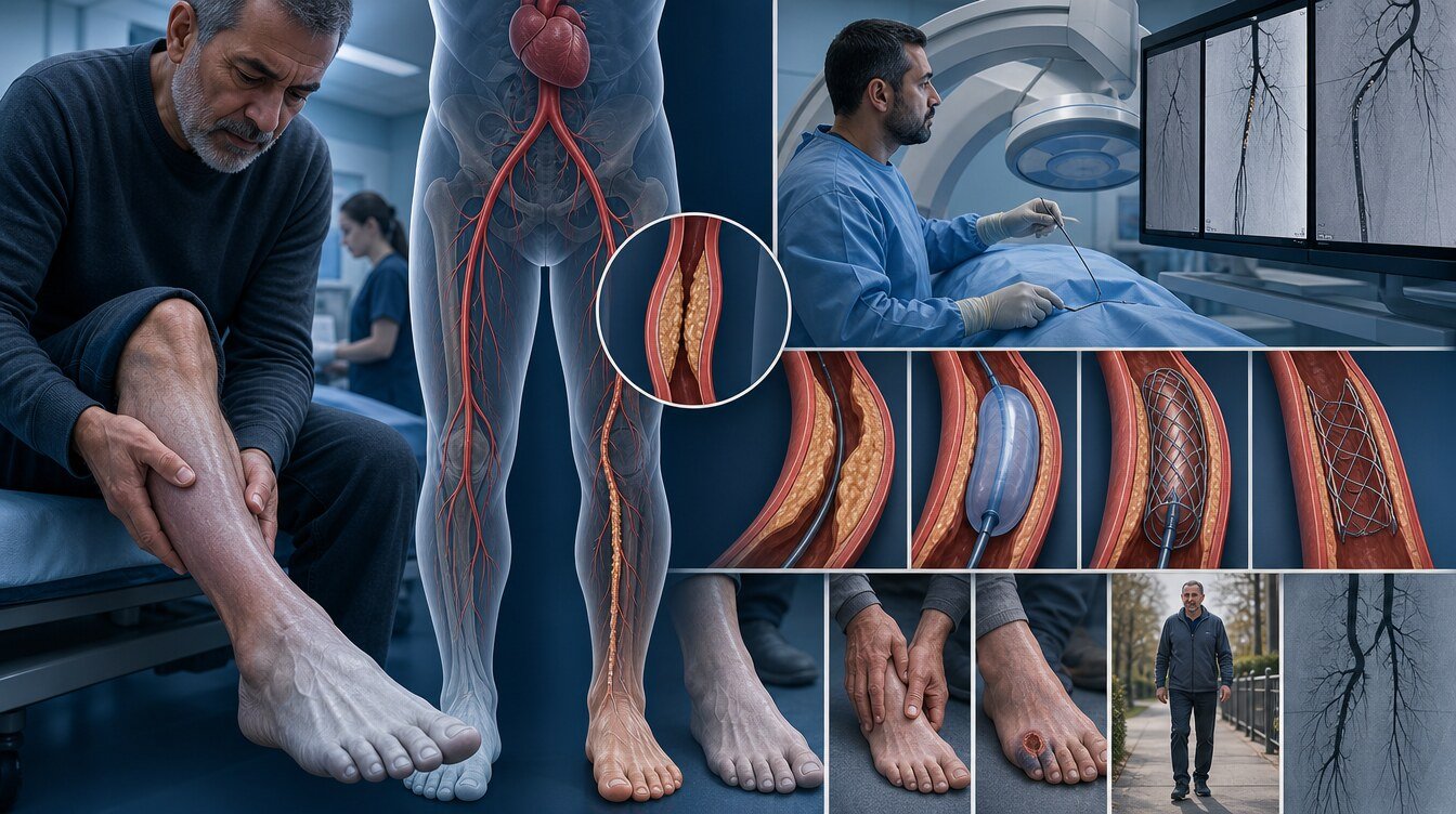
After a straightforward endovascular procedure a short observation may be enough; in CLTI or complex anatomy the stay may be longer. The access site should be kept clean, heavy lifting avoided in the first days, walking started gradually, and medicines and reviews not missed.
The feet should be checked every day for colour change, blistering and wounds. Bare feet, tight shoes and direct heat are especially risky when feeling is reduced. Even a wound that looks small should be shown without delay in PAD or diabetes.[1,13] Follow-up may use examination, ABI/TBI or duplex. Restenosis or progression in another segment may lead to a further procedure; repeating the same procedure is not always the most suitable option.[1,2]
Emergency symptoms
Sudden severe leg or foot pain, a cold or pale foot, new numbness or weakness, inability to move the foot, bleeding from the access site that will not stop, a groin swelling that grows quickly, fever and discharge around a wound, new blackening, chest pain or fainting should be assessed without waiting for a planned review. Sudden pain, coldness, paleness, numbness and loss of strength together suggest acute limb ischaemia.[1,2]
Academic context of Prof. Dr. Hakan Uçar
Among the sources that could be verified for this package, no confirmed peripheral artery disease, peripheral stenting, CLTI or limb-ischaemia outcome paper by Prof. Dr. Hakan Uçar was found. No specific scientific paper, method or success rate in PAD is therefore attributed to him. Coronary, carotid or TAVI papers are not used as PAD evidence.
Frequently asked questions
No. PAD is narrowing of arteries and reduced blood flow to the tissues of the leg. Varicose veins and venous insufficiency are disorders of blood returning to the heart. Symptoms can look similar, so examination is needed.
A sudden loss of blood flow can cause lasting nerve and muscle injury within hours. Sudden pain, coldness, paleness, numbness or loss of strength needs emergency assessment.[1]
No single duration can be given. The arterial segment, lesion length, calcification, diabetes, smoking, the device and medicine adherence affect patency. Regular follow-up helps detect re-narrowing earlier.
In most people, walking at the time and pace advised by the care team matters. An open wound, infection, acute ischaemia or a specific post-procedure restriction leads to a different plan.[7]
No. Better flow can help wound healing; infection, pressure, diabetes and wound depth also matter. Patency is not the same as wound healing.[13]
In a person with PAD, diabetes or loss of feeling, even a small wound matters. Redness, discharge, a bad smell, blackening or failure to heal should be assessed without delay.[13]
Literature
References
Gornik HL, Aronow HD, Goodney PP, et al. 2024 ACC/AHA/AACVPR/APMA/ABC/SCAI/SVM/SVN/SVS/SIR/VESS Guideline for the Management of Lower Extremity Peripheral Artery Disease. Circulation. 2024;149:e1313–e1410. DOI: 10.1161/CIR.0000000000001251
Mazzolai L, Teixidó-Turà G, Lanzi S, et al. 2024 ESC Guidelines for the management of peripheral arterial and aortic diseases. European Heart Journal. 2024;45:3538–3700. DOI: 10.1093/eurheartj/ehae179
Farber A, Menard MT, Conte MS, et al. Surgery or Endovascular Therapy for Chronic Limb-Threatening Ischemia. New England Journal of Medicine. 2022;387:2305–2316. DOI: 10.1056/NEJMoa2207899
Bradbury AW, Moakes CA, Popplewell M, et al. A vein bypass first versus a best endovascular treatment first revascularisation strategy for patients with chronic limb threatening ischaemia (BASIL-2). The Lancet. 2023;401:1798–1809. DOI: 10.1016/S0140-6736(23)00462-2
Bonaca MP, Bauersachs R, Anand SS, et al. Rivaroxaban in Peripheral Artery Disease after Revascularization. New England Journal of Medicine. 2020;382:1994–2004. DOI: 10.1056/NEJMoa2000052
Anand SS, Bosch J, Eikelboom JW, et al. Rivaroxaban with or without aspirin in patients with stable peripheral or carotid artery disease. The Lancet. 2018;391:219–229. DOI: 10.1016/S0140-6736(17)32409-1
Hageman D, Fokkenrood HJP, Gommans LNM, van den Houten MML, Teijink JAW. Supervised exercise therapy versus home-based exercise therapy versus walking advice for intermittent claudication. Cochrane Database of Systematic Reviews. 2018;(4):CD005263. DOI: 10.1002/14651858.CD005263.pub4
Nordanstig J, James S, Andersson M, et al. Mortality with Paclitaxel-Coated Devices in Peripheral Artery Disease. New England Journal of Medicine. 2020;383:2538–2546. DOI: 10.1056/NEJMoa2005206
Parikh SA, Schneider PA, Mullin CM, et al. Mortality in randomised controlled trials using paclitaxel-coated devices for femoropopliteal interventional procedures: an updated patient-level meta-analysis. The Lancet. 2023;402:1848–1856. DOI: 10.1016/S0140-6736(23)02189-X
Falkenberg M, James S, Andersson M, et al. Paclitaxel-coated versus uncoated devices for infrainguinal endovascular revascularisation in chronic limb-threatening ischaemia (SWEDEPAD 1). The Lancet. 2025;406:1103–1114. DOI: 10.1016/S0140-6736(25)01585-5
Nordanstig J, James S, Andersson M, et al. Paclitaxel-coated versus uncoated devices for infrainguinal endovascular revascularisation in patients with intermittent claudication (SWEDEPAD 2). The Lancet. 2025;406:1115–1127. DOI: 10.1016/S0140-6736(25)01584-3
Bausback Y, Wittig T, Schmidt A, et al. Drug-Eluting Stent Versus Drug-Coated Balloon Revascularization in Patients With Femoropopliteal Arterial Disease (REAL PTX). Journal of the American College of Cardiology. 2019;73:667–679. DOI: 10.1016/j.jacc.2018.11.039
Zhan LX, Branco BC, Armstrong DG, Mills JL. The Society for Vascular Surgery lower extremity threatened limb classification system based on Wound, Ischemia, and foot Infection (WIfI) correlates with risk of major amputation and time to wound healing. Journal of Vascular Surgery. 2015;61:939–944. DOI: 10.1016/j.jvs.2014.11.045
This content is for general information only and does not replace personalized medical advice. In emergencies call local emergency services.
Continue