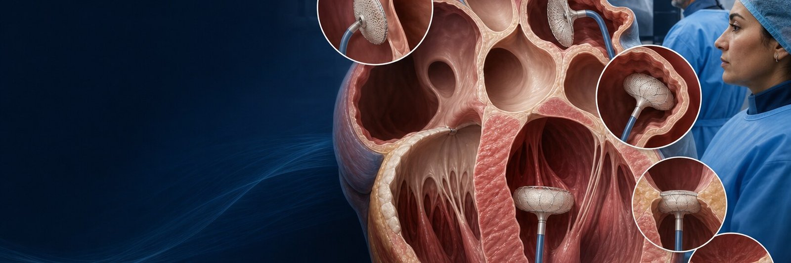
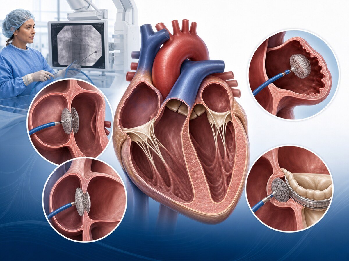
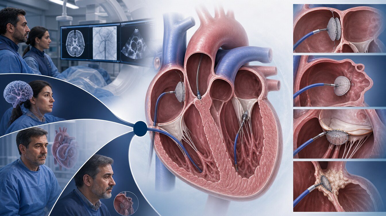
Structural heart closure procedures close a congenital or acquired opening, leak or clot-prone region between the heart chambers or around a heart valve through a catheter, a thin tube advanced through a blood vessel. These procedures are not for everyone. Suitable candidates are chosen by the type of disease, symptoms, heart anatomy, clot or stroke risk, bleeding risk and the other treatment options. The decision is often made by a Structural Heart Team.
This text is general patient education; it is not a diagnosis or a personal procedure decision. It requires a final physician review before publication.
Which four procedures does this parent page cover?
| Procedure | What is closed? | Dedicated page |
|---|---|---|
| PFO closure | A small atrial passage that did not close fully after birth | PFO Closure |
| ASD closure | A congenital tissue defect between the atria | ASD Closure |
| LAA closure | The clot-prone appendage of the left atrium | LAA Closure |
| PVL closure | A leak around a prosthetic heart valve | PVL Closure |
Randomised evidence from one procedure is not transferred to another. The antithrombotic plan also differs; one aspirin, antiplatelet or anticoagulant scheme cannot be applied to all four.
What are structural heart disease and closure?
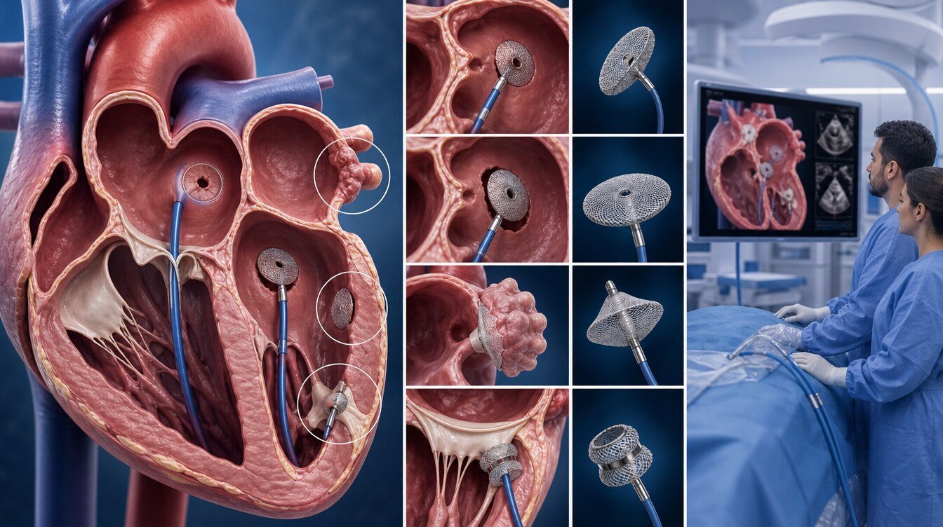
Structural heart disease is the general name for anatomical problems in the heart chambers, valves, walls or the great vessels attached to the heart. Some are congenital; some appear after valve surgery or valve implantation.
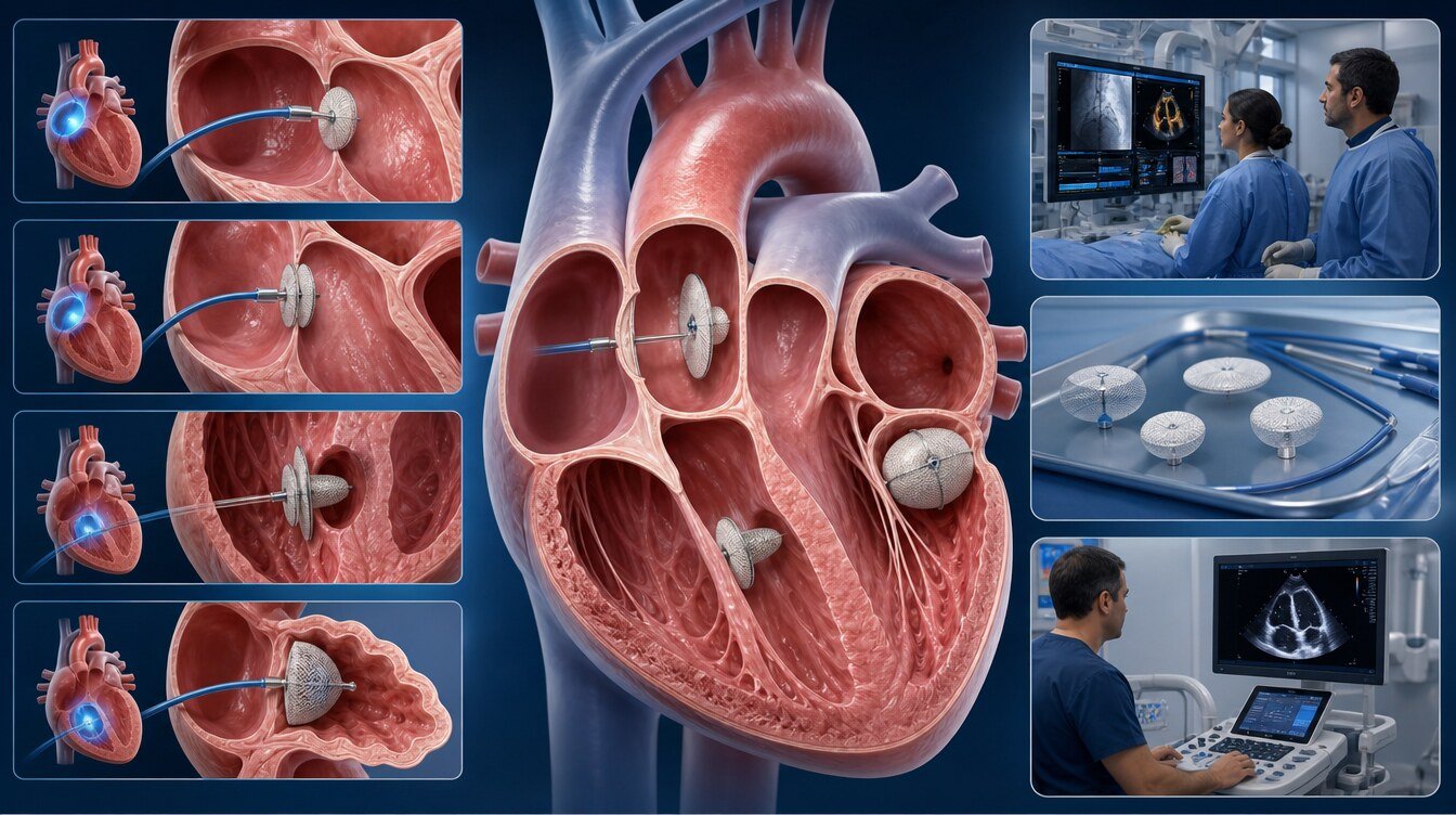
Percutaneous closure usually places a closure system at the target through catheters from a groin vein or artery. X-ray imaging and echocardiography are used during the procedure. Once the position is confirmed the system is released and, over time, becomes incorporated into the heart tissue.
Catheter repair or replacement of heart valves is not the subject of this page; that cluster sits under heart valve interventions.
The role of the Structural Heart Team
The Structural Heart Team may include an interventional cardiologist, a cardiac surgeon, a cardiac imaging specialist and, when needed, a neurologist, an electrophysiologist, an adult congenital-heart specialist, an anaesthetist and other clinicians.
The core questions are:
- Does this problem truly need to be closed?
- Is catheter closure anatomically possible?
- Could open or closed surgery be safer or more effective?
- Is medical therapy and surveillance enough?
- What is the balance of benefit and bleeding risk from medicines after the procedure?
Tests used in the assessment
- TTE (transthoracic echocardiography): standard ultrasound of the heart from the chest wall.
- TEE (transoesophageal echocardiography): more detailed imaging with a probe in the oesophagus. It is used before closure to assess the opening, the valve or the shape of the LAA.
- Bubble study: a special echocardiogram that shows whether small air bubbles given into a vein pass from the right side of the heart to the left. It can be used when a PFO is being investigated.
- Cardiac CT: cross-sectional images of the heart and vessels. It can help plan complex ASD, LAA and PVL anatomy.
- Rhythm monitoring: ECG, Holter or longer recording. It is used to look for a rhythm-related stroke cause when a PFO is found, and to document atrial fibrillation when LAA closure is considered.
- Stroke work-up: brain imaging, vessel studies, rhythm monitoring and a search for other causes are needed when a PFO is found. Finding a PFO by chance does not mean the PFO caused the stroke.
- Dental and infection review: an active dental abscess, a febrile infection or suspected endocarditis can delay the procedure. This review matters especially in people with a prosthetic valve and PVL.
General preparation and the procedure day
Preparation follows the structure that will be closed. Current illnesses, operations, allergies, bleeding history and all medicines are reviewed. Blood thinners, antiplatelet medicines, diabetes medicines and herbal products should be reported. No medicine should be stopped without the team’s advice.
Blood counts, kidney function, clotting tests, blood group, ECG and a lung review may be requested. In ASD, pulmonary vascular resistance and the load on the right heart are assessed; in LAA, shape, depth and neighbouring structures; in PVL, the site and size of the leak and how the prosthetic valve is working.
On the day, follow the fasting advice; do not stop medicines unless told; bring identification, test results and a medicine list; and arrange a companion because you will not drive afterwards.
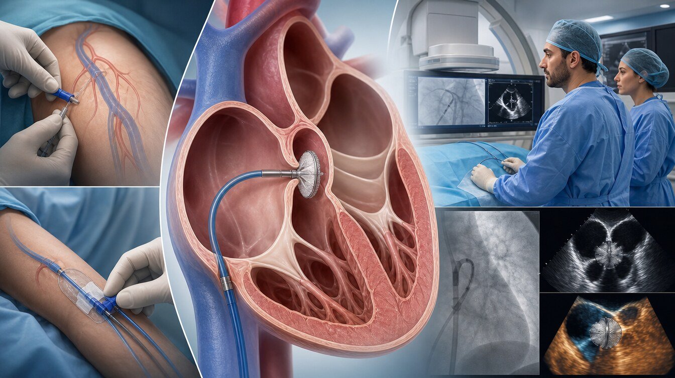
Anaesthesia depends on the procedure. Some are done with conscious sedation; longer or TEE-guided procedures may use general anaesthesia. A catheter is advanced from the groin; TEE, TTE, intracardiac echocardiography and fluoroscopy assess the target; the system is released if the position is confirmed. Duration depends on anatomy. Some people go home the same day; others stay overnight.
Recovery and follow-up
Mild groin pain or bruising can occur after discharge. Avoid heavy lifting and strenuous activity in the first days. Keep the access site clean; growing swelling, active bleeding or expanding bruising should lead to review.
Antithrombotic treatment is not the same after every procedure. The type and duration of antiplatelet or anticoagulant therapy are individualised to the structure closed, the system used, rhythm disease, bleeding risk, kidney function and other illnesses.
Follow-up may include ECG, TTE or TEE when needed. The aims are to check the position of the system, residual opening or leak, the heart chambers and possible rhythm problems.
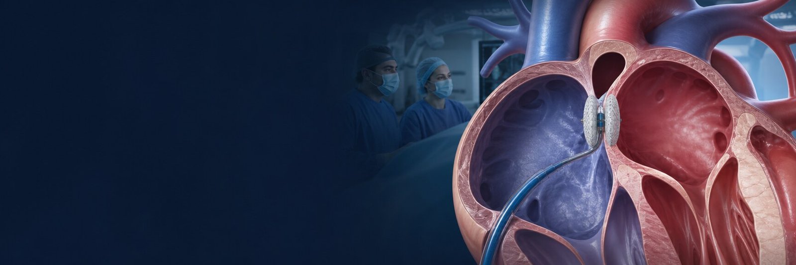
PFO closure
A PFO is a small passage between the atria that is needed before birth and stays open in some people. It is common and causes no disease in most people. Finding a PFO is not by itself a reason for closure.
In some people a clot from a vein can pass from right to left and contribute to paradoxical embolism. Other causes of stroke must be excluded before a causal link is assumed.
Closure may be considered in carefully selected people aged 18–60 who have had an ischaemic stroke of unexplained cause after a thorough work-up. A large right-to-left shunt and an atrial septal aneurysm may raise the chance of benefit. The 2024 ESO guideline supports closure plus antiplatelet therapy in selected PFO-related stroke; it advises against closure when the PFO appears unrelated to the stroke.[1]
RESPECT, CLOSE, REDUCE, DEFENSE-PFO and meta-analyses suggest that recurrent stroke risk can fall versus medicine in selected people, while new atrial fibrillation can increase. In CLOSURE I, closure was not superior to medicine for preventing stroke or transient ischaemic attack. Results are not generalised to every incidental PFO.[2–5]
After PFO closure the choice and duration of antiplatelet therapy are individual. An anticoagulant is not automatically required in every person unless there is another medical reason. Detail belongs on the PFO Closure page.
ASD closure
An ASD is a congenital opening in the wall between the atria. The most common type, secundum ASD, usually causes a left-to-right shunt. Over time the right atrium and right ventricle can enlarge. If pulmonary vascular resistance rises, the closure decision becomes more complex. Closure can be harmful in Eisenmenger physiology.
In adults, secundum ASD closure may be considered when the right heart is enlarged and the left-to-right shunt is meaningful, with or without symptoms. When the anatomy is suitable, catheter closure is often one of the preferred options. Most of the evidence is observational rather than randomised. An AHA/ACC systematic review found a signal of better functional status and right-ventricular size and pressure; evidence was insufficient to determine an effect on death.[6]
Primum ASD and sinus venosus ASD have different anatomy. Valve problems or anomalous pulmonary venous connection may accompany them, so surgery is usually needed. Not every ASD can be closed by catheter. Results from PFO or LAA trials are not transferred to ASD.
Detail belongs on the ASD Closure page.
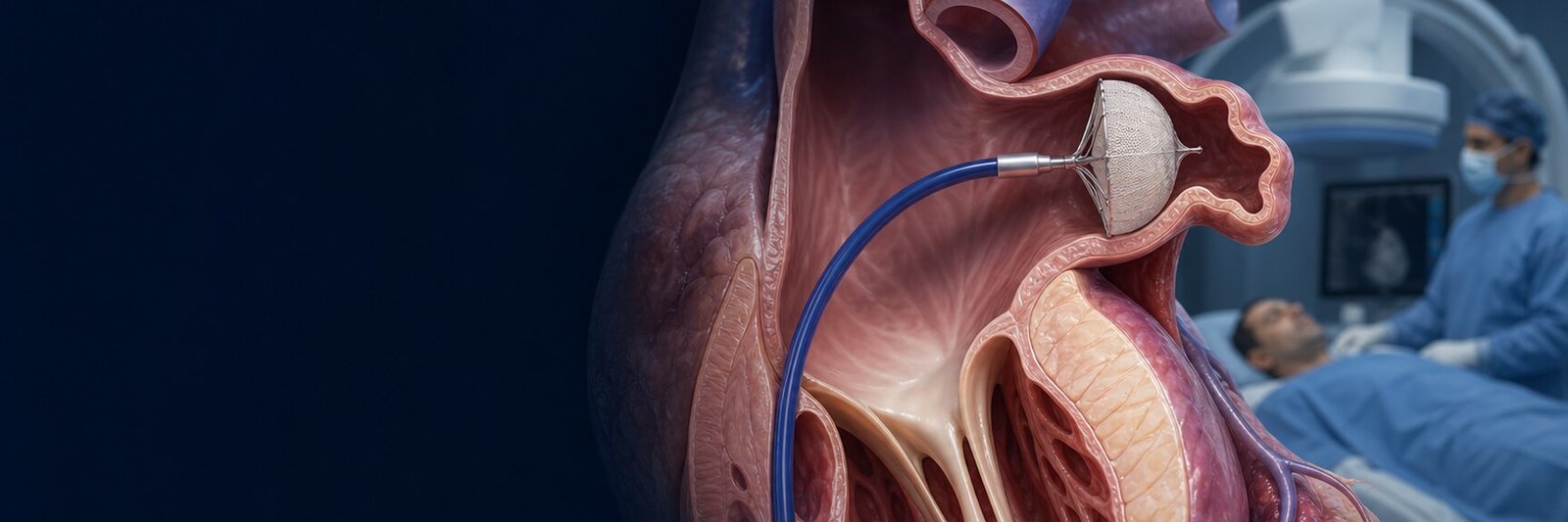
LAA closure
The left atrial appendage is a heart outpouching in which blood can stagnate and clot during atrial fibrillation. LAA closure is an option to reduce heart-origin stroke risk in people with nonvalvular atrial fibrillation. The procedure does not abolish atrial fibrillation and does not prevent every cause of stroke.
DOACs and warfarin remain effective medical options. LAA closure is not an automatic replacement for everyone. PROTECT AF and PREVAIL compared LAA closure with warfarin. In PRAGUE-17, in people at high stroke and bleeding risk, LAA closure met a “not worse” criterion versus DOAC for preventing major cardiovascular, neurological or bleeding events; the finding was kept at four years, with fewer non-procedure bleeds. These results are not a superiority guarantee for every person.[7–10]
The post-procedure antithrombotic protocol can vary with the system and with bleeding and stroke risk. There is no single universal regimen. Follow-up looks for device-related thrombus and residual leak. Detail belongs on the LAA Closure page.
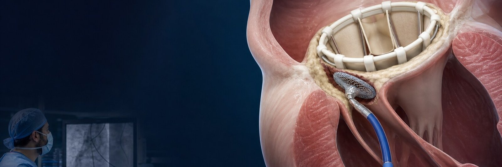
PVL closure
A PVL is blood leaking around a surgical or some transcatheter prosthetic valves. Some PVLs cause no symptoms; others can lead to heart failure or haemolysis.[11,12]
TEE, especially three-dimensional TEE, is important for locating and sizing the leak. Endocarditis must be excluded first. Catheter PVL closure is usually not appropriate when infection is active.
When a PVL causes symptoms, heart failure or haemolysis, repeat surgery is compared with catheter repair. Technical success (reducing the leak) and clinical benefit (relief of breathlessness, heart failure or haemolysis) are separate outcomes. PVL evidence is mostly from registries and observational series; randomised comparisons are limited.[11,12]
Detail belongs on the PVL Closure page.
A four-branch decision path
The frame below does not replace a personal decision.
- Structural heart assessment starts; the core problem is separated as PFO, ASD, LAA or PVL.
- If PFO: selected cryptogenic stroke with no other cause uses the frame on the PFO Closure page.
- If ASD: secundum ASD and right-heart enlargement are reviewed on ASD Closure; primum and sinus venosus are kept separate.
- If LAA: nonvalvular AF and stroke prevention belong on LAA Closure; it is not rhythm therapy.
- If PVL: a meaningful leak around a prosthetic valve, exclusion of infection and comparison with surgery belong on PVL Closure.
- Shared decision-making, a procedure-specific antithrombotic plan and scheduled imaging follow.
Shared risks and procedure-specific emphasis
Risks vary with the disease, the anatomy and general health:
- Vessel injury, bleeding and haematoma
- Pericardial effusion; tamponade is considered especially in LAA and some ASD procedures
- Device embolisation
- Residual shunt or leak
- Arrhythmia; new atrial fibrillation is part of the PFO decision, and rhythm disease is already the diagnosis in LAA
- Stroke
- Device thrombus, watched especially after LAA closure
- Infection; active endocarditis delays PVL closure
- Contrast reaction and kidney injury
None of these risks has the same likelihood in every person.
Who may not be suitable, and what are the alternatives?
- PFO: Closure may not be suitable if another stroke cause is found, the PFO looks incidental or there is active infection. Alternatives are antiplatelet therapy, anticoagulation in selected people and treatment of other stroke risk factors.
- ASD: Closure can be unwise if there is not enough rim, pulmonary vascular resistance is very high or Eisenmenger physiology is present. Alternatives are surveillance, medicines for symptoms and surgery.
- LAA: Clot inside the LAA, unsuitable anatomy, active infection or inability to take even short-term antithrombotic therapy can block the procedure. Alternatives are a DOAC, warfarin and rate or rhythm control.
- PVL: Active endocarditis, a seriously malfunctioning prosthesis or a leak that cannot be targeted by catheter need surgery or another treatment.
Emergency warnings after a procedure
Seek emergency help if any of the following is severe or increasing quickly:
- New or worsening chest pain
- Breathlessness at rest, fainting or bluish colour
- New marked palpitations or an irregular heartbeat
- Facial droop, weakness in an arm or leg, slurred speech or sudden loss of vision
- Fever, shivering or a sudden decline
- Uncontrolled bleeding or rapidly growing swelling at the groin
- Sudden weakness, pallor or dark urine
Questions for a Structural Heart Team
- Which features of PFO, ASD, LAA or PVL does my diagnosis have?
- What is the medical reason for closure, and what is the risk of not doing a procedure?
- Which other stroke, rhythm or infection causes have been excluded?
- Is my anatomy suitable for a catheter procedure?
- What are the alternatives of surgery, medicine or surveillance?
- Which anaesthesia will be used?
- How long will I take a blood thinner or antiplatelet medicine?
- When will follow-up TTE, TEE, CT or rhythm monitoring be done?
- What happens if device thrombus, residual shunt or arrhythmia is found?
- Which symptoms should send me to emergency care?
How the evidence should be read
The evidence on this page is not all of the same strength:
- Guideline recommendation: ESO 2024 belongs here for selected people with PFO-related stroke.[1]
- Randomised trial: important randomised trials exist for PFO and LAA.[2,3,9]
- Meta-analysis: PFO meta-analyses show less stroke together with more atrial fibrillation.[4]
- Registry or observational study: much of the ASD and PVL evidence is of this type; uncertainty about causation is greater.[6,11,12]
A favourable trial result does not mean the same benefit for every person.
Academic context of Prof. Dr. Hakan Uçar
Among the sources that could be verified for this package, no confirmed PFO, ASD, LAA or PVL closure outcome paper by Prof. Dr. Hakan Uçar is used as evidence on this parent page. No specific procedure volume, device or success rate is attributed to him.
Frequently asked questions
No. The type and size of the opening, its effect on blood flow, changes in the heart chambers and the person’s symptoms are considered together.
No. A PFO is a passage that did not close fully after birth; an ASD is a true tissue defect between the atria. Diagnosis, the reason for closure and follow-up differ.
Usually not. Closure is not usually chosen in a person who has not had a stroke or whose stroke has another cause.[1]
Right-heart enlargement and symptoms can improve, but recovery varies with age, the timing of closure, pulmonary pressure and accompanying rhythm problems.[6]
Closing the defect in advanced pulmonary vascular disease can disturb the balance between the heart and the lung circulation. Pulmonary vascular resistance is therefore reviewed first.
No. LAA closure does not correct atrial fibrillation; in selected people it aims to reduce stroke risk related to that rhythm.[9]
Not always. Short- or longer-term treatment follows device follow-up, clot risk, bleeding risk and other diseases. There is no single universal regimen.
It can follow loosening, calcification, tissue injury or infection around a prosthetic valve. Not every PVL causes symptoms.[11]
Catheter closure is not open surgery. In some anatomies surgery can still be more suitable or safer than a catheter method.
Some procedures use sedation and local anaesthesia; others use general anaesthesia. The choice follows complexity, the need for TEE and general health.
Most closure systems are left in the heart and become incorporated into the tissue over time. Position and nearby effects are checked on follow-up imaging.
Most people return gradually to usual life; the time to heavy lifting and intense exercise depends on the procedure, the access site and the control results.
Residual shunt or leak, clot on the device, rhythm disturbance, valve function and the response of the heart chambers can change over time. Planned reviews therefore matter.
Literature
References
Caso V, Turc G, Abdul-Rahim AH, et al. European Stroke Organisation (ESO) guidelines on the diagnosis and management of patent foramen ovale (PFO) after stroke. European Stroke Journal. 2024;9:800–834. DOI: 10.1177/23969873241247978
Søndergaard L, Kasner SE, Rhodes JF, et al. Patent Foramen Ovale Closure or Antiplatelet Therapy for Cryptogenic Stroke. New England Journal of Medicine. 2017;377:1033–1042. DOI: 10.1056/NEJMoa1707404
Carroll JD, Saver JL, Thaler DE, et al. Closure of Patent Foramen Ovale versus Medical Therapy after Cryptogenic Stroke. New England Journal of Medicine. 2013;368:1092–1100. DOI: 10.1056/NEJMoa1301440
Ahmad Y, Howard JP, Arnold A, et al. Patent foramen ovale closure vs. medical therapy for cryptogenic stroke: a meta-analysis of randomized controlled trials. European Heart Journal. 2018;39:1638–1649. DOI: 10.1093/eurheartj/ehy121
Lee PH, Song J-K, Kim JS, et al. Cryptogenic Stroke and High-Risk Patent Foramen Ovale. Journal of the American College of Cardiology. 2018;71:2335–2342. DOI: 10.1016/j.jacc.2018.02.046
Oster ME, Bhatt AB, Zaragoza-Macias E, Dendukuri N, Marelli A. Interventional Therapy Versus Medical Therapy for Secundum Atrial Septal Defect: A Systematic Review (Part 2) for the 2018 AHA/ACC Guideline. Journal of the American College of Cardiology. 2019;73:1579–1595. DOI: 10.1016/j.jacc.2018.08.1032
Holmes DR, Kar S, Price MJ, et al. Prospective Randomized Evaluation of Left Atrial Appendage Closure Versus Long-Term Warfarin Therapy. Journal of the American College of Cardiology. 2014;64:1–12. DOI: 10.1016/j.jacc.2014.04.029
Reddy VY, Sievert H, Halperin J, et al. Percutaneous Left Atrial Appendage Closure vs Warfarin for Atrial Fibrillation. JAMA. 2014;312:1988–1998. DOI: 10.1001/jama.2014.15192
Osmancik P, Herman D, Neuzil P, et al. Left Atrial Appendage Closure Versus Direct Oral Anticoagulants in High-Risk Patients With Atrial Fibrillation. Journal of the American College of Cardiology. 2020;75:3122–3135. DOI: 10.1016/j.jacc.2020.04.067
Osmancik P, Herman D, Neuzil P, et al. 4-Year Outcomes After Left Atrial Appendage Closure Versus Nonwarfarin Oral Anticoagulation for Atrial Fibrillation. Journal of the American College of Cardiology. 2022;79:1–14. DOI: 10.1016/j.jacc.2021.10.023
Rihal CS, Sorajja P, Booker JD, Hagler DJ, Cabalka AK. Principles of Percutaneous Paravalvular Leak Closure. JACC: Cardiovascular Interventions. 2012;5:121–130. DOI: 10.1016/j.jcin.2011.11.007
Millán X, Skaf S, Joseph L, et al. Transcatheter Reduction of Paravalvular Leaks: A Systematic Review and Meta-analysis. Canadian Journal of Cardiology. 2015;31:260–269. DOI: 10.1016/j.cjca.2014.12.012
This content is for general information only and does not replace personalized medical advice. In emergencies call local emergency services.
Continue