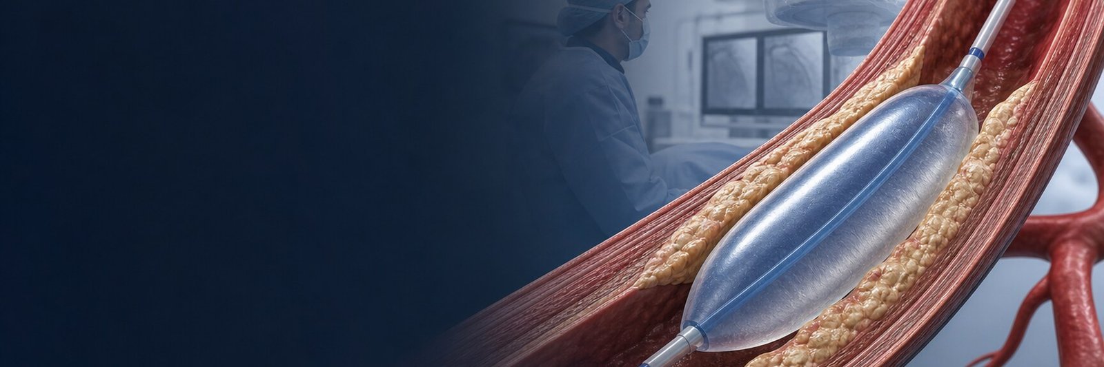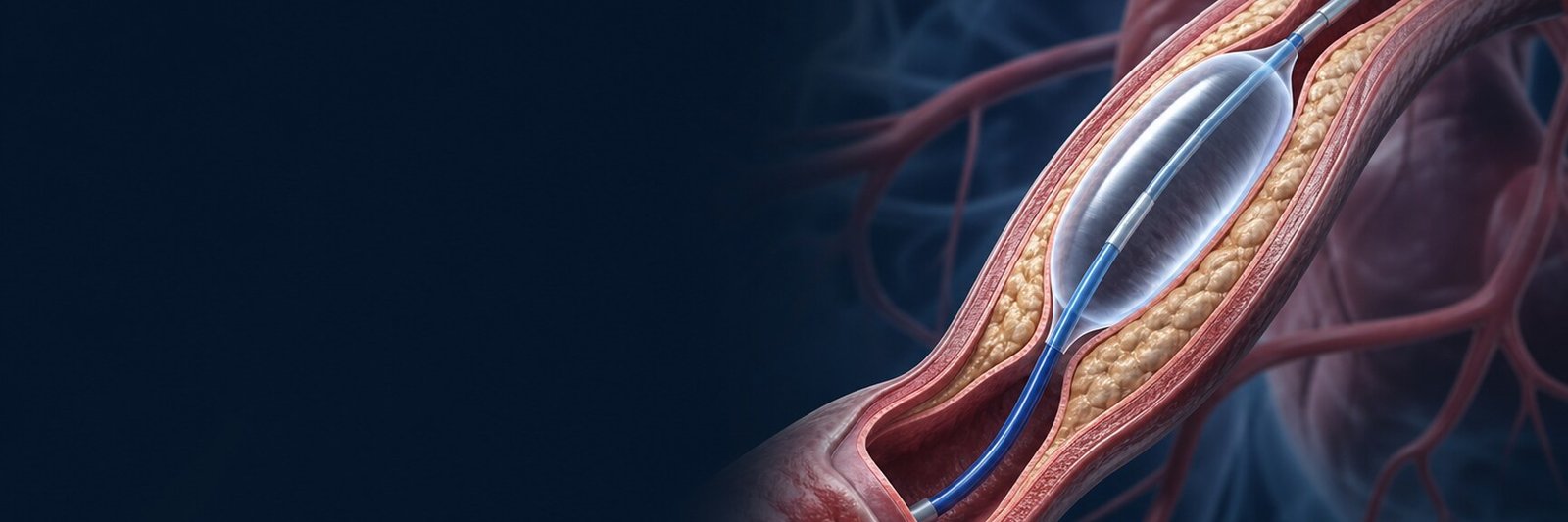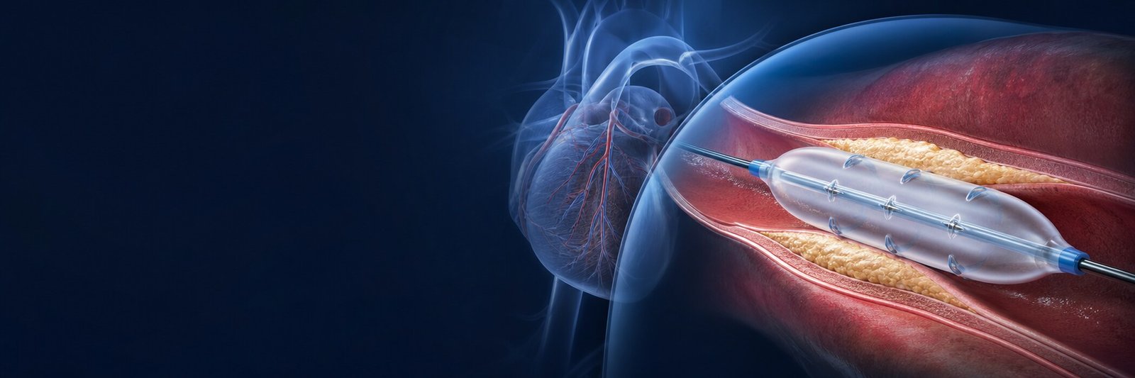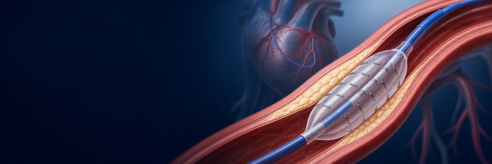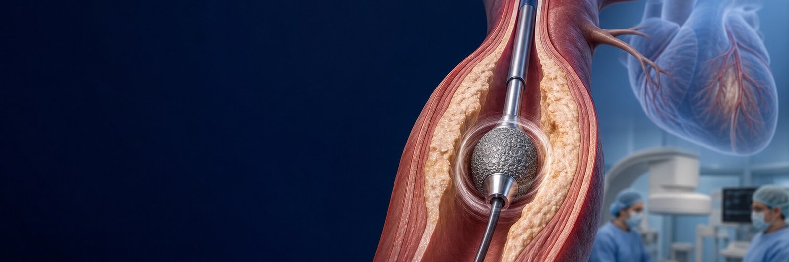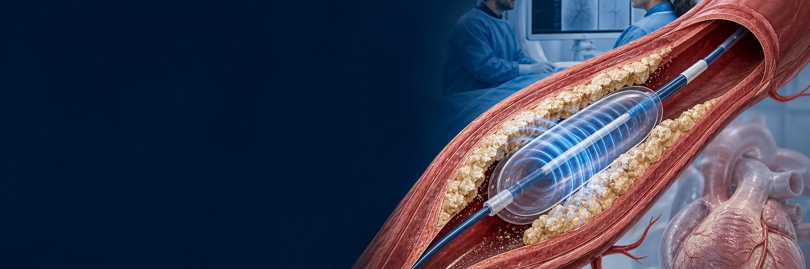
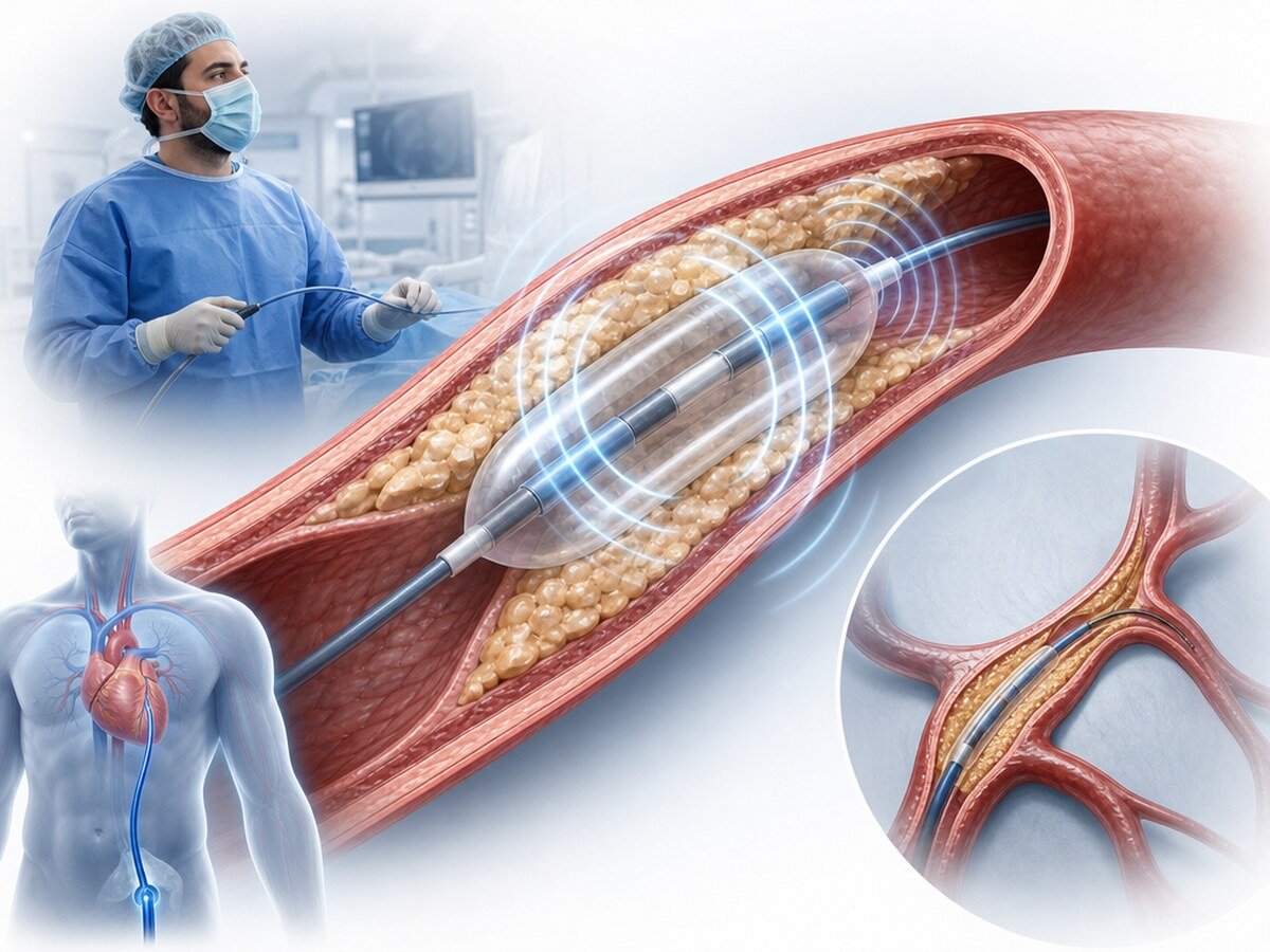
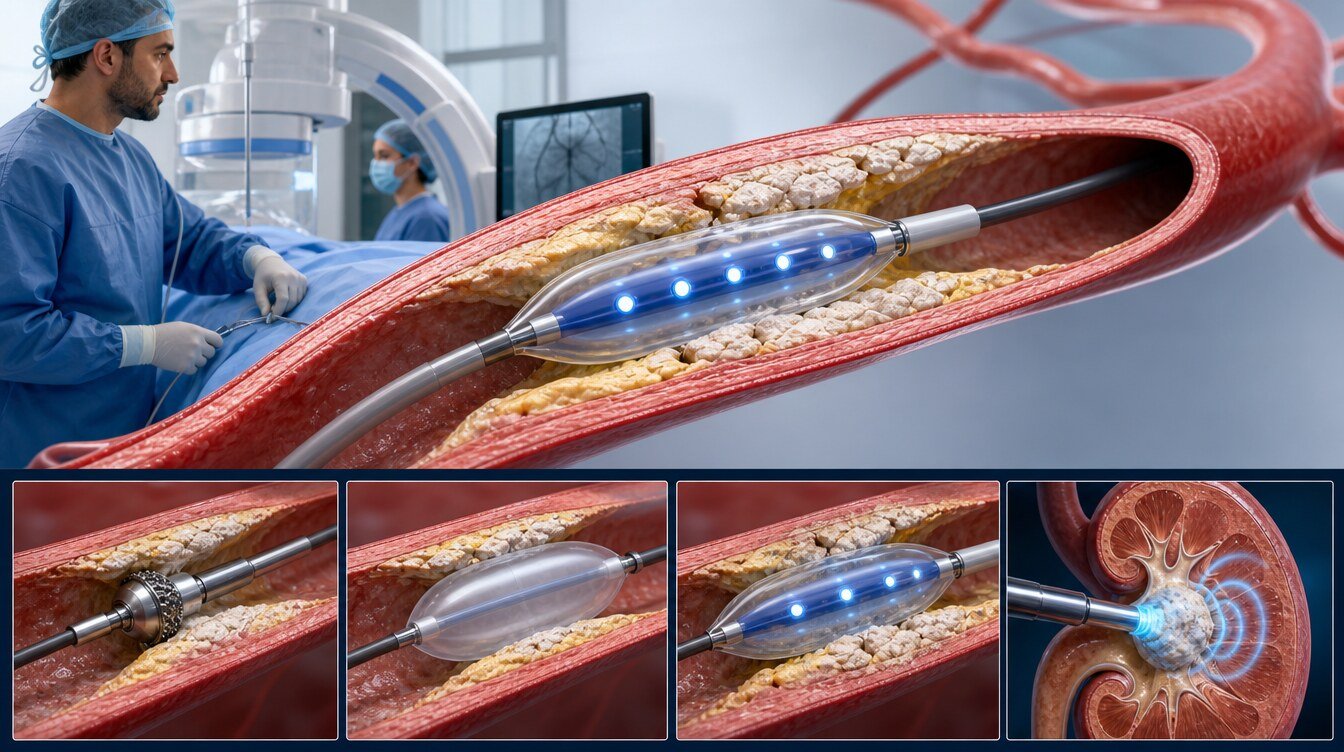
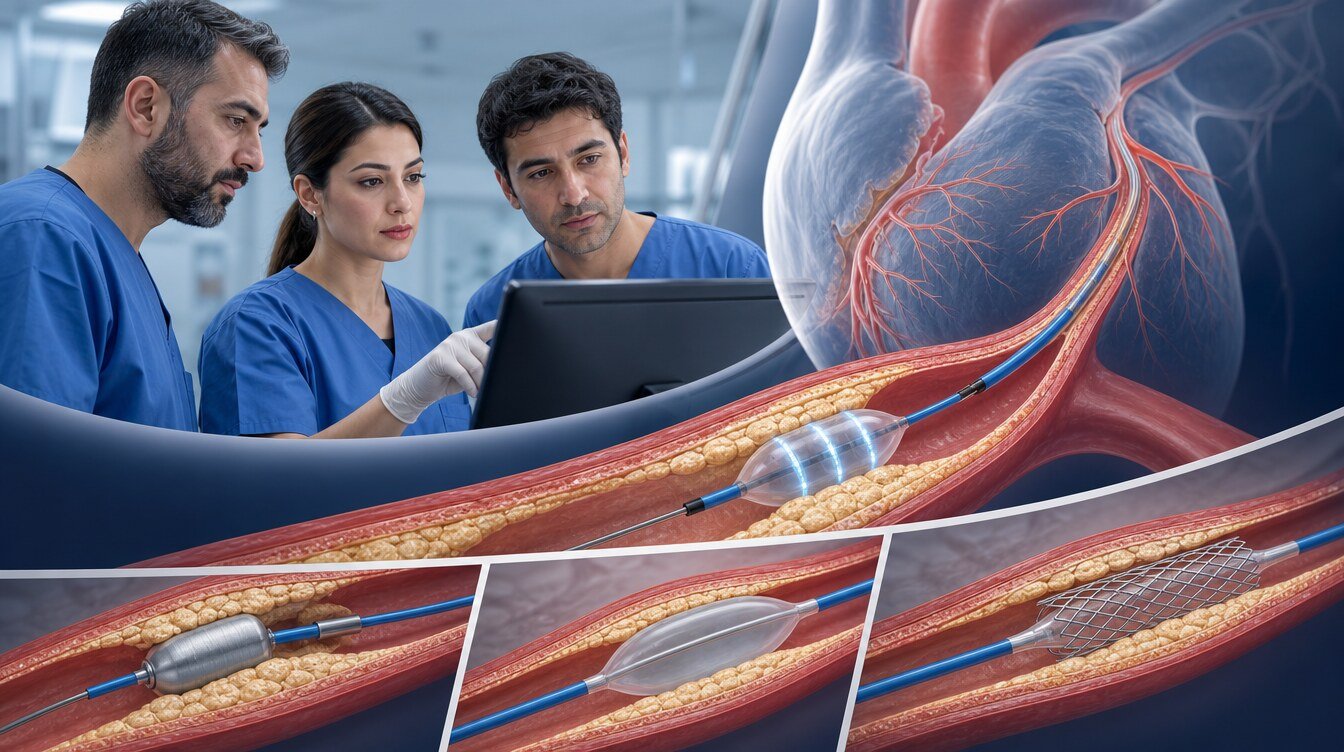
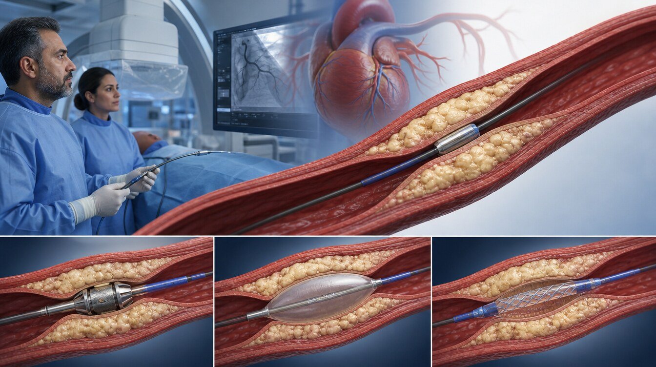
Coronary intravascular lithotripsy (IVL) is a temporary intravascular tool used to prepare a hard, calcified coronary stenosis for a stent or another treatment. A dedicated balloon advanced over a fine guidewire delivers brief acoustic pressure waves intended to change the mechanical behaviour of calcium. The catheter is removed afterwards and is not left in the artery. It is not required in every calcified stenosis, and calcium is not cleared from the body.[3,22,26]
What does IVL mean?
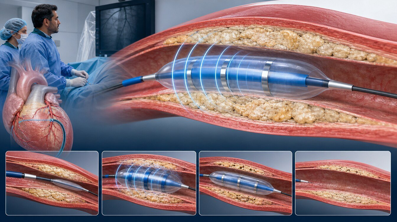
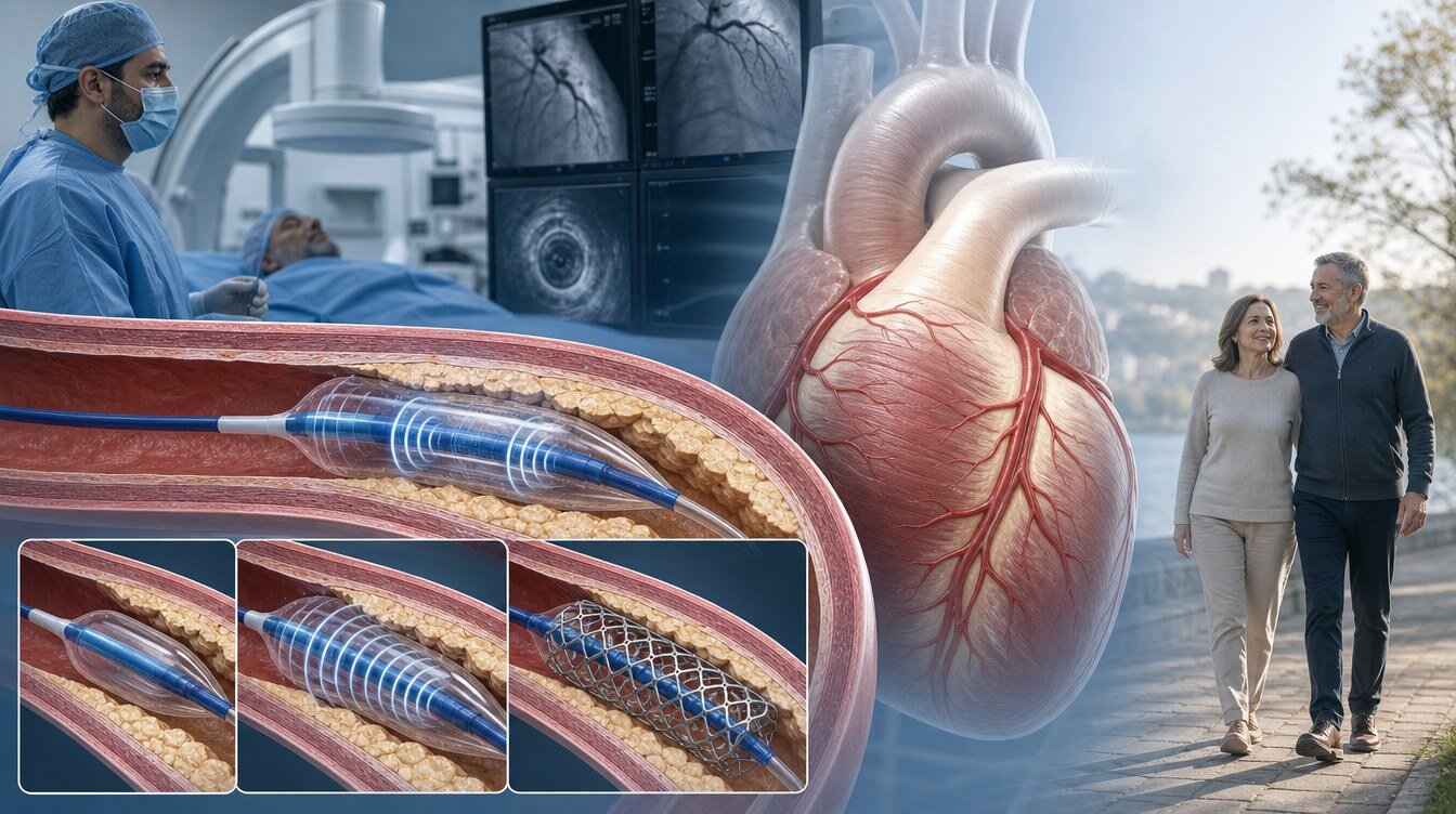
Coronary IVL is a calcium-modification method that uses a temporary balloon catheter placed inside the artery. The aim is to create fractures in calcium in a selected stenosis so that the vessel wall becomes more compliant and a stent can open more controllably. That does not mean calcium is destroyed, vaporised or cleared from the artery. Fractures are not guaranteed in every lesion.[2,3,26]
In everyday language, “intravascular shockwave” describes the acoustic pressure waves delivered by the balloon. It is not extracorporeal kidney-stone lithotripsy. Coronary IVL is performed from inside the artery during angioplasty in a cardiac catheter laboratory.[3,26]
Balloon diameter, pressure, pulse count and inflation cycles are device- and lesion-specific. This page therefore does not publish a universal size, pressure or pulse recipe. IVL is not a permanent implant like a stent; after preparation the balloon is withdrawn with the catheter.[26]
What do Shockwave and ShockFast mean?
Shockwave is a brand and platform name; for example the C2 coronary IVL catheter named in studies belongs to this platform. The generic medical name of the procedure is intravascular lithotripsy (IVL). The brand name is not used as a menu label, heading or “standard treatment for everyone”.[26]
ShockFast is not a misspelling of that name. Records list it as a separate coronary IVL device from Shunmei Medical, designed for comparison with Shockwave IVL. At the cut-off date the trial record had not published results; ShockFast must not be presented as a generic IVL synonym or as a proven superior device.[21]
When may it be considered?
Coronary IVL is not a routine step in every calcified stenosis. Use depends on whether the balloon can cross the stenosis, calcium depth and circumference, the stent plan, imaging findings, and operator and centre experience.[22,23]
Severe calcified new (de novo) stenoses
The DISRUPT CAD studies and most randomised evidence examine pre-stent preparation in severely calcified new (de novo) coronary stenoses. That is the strongest direct evidence setting; it does not transfer automatically to every anatomy or clinical picture.[3,5,14]
Calcium phenotype selected with imaging
IVUS or OCT can help distinguish whether calcium is superficial or deep and how far it encircles the vessel. Phenotype may influence the choice among IVL, balloons, rotational atherectomy or a combined strategy; an image alone does not mandate the method.[22,23]
Tissue inside a stent or an under-expanded stent (a different question)
Registries and case series exist for underexpansion related to calcium beneath a stent or for in-stent narrowing. That use may vary by indication and country; it is not the same as the de novo pivotal studies and is not written here as a standard, universally approved indication.[22,23]
When may it be unsuitable or insufficient alone?
The IVL balloon must still cross the target stenosis. Lesions that the balloon cannot cross, that are highly tortuous, or that the device cannot reach may need another preparation first. That does not mean one tool is universally superior.[22,23]
Thrombotic, freshly occlusive lesions (for example selected ACS/STEMI) were often excluded from the pivotal device studies. Broad claims of superiority in that group are not supported; the decision is individual.[3,22]
A different strategy may be needed when:
- The IVL balloon cannot cross the stenosis safely
- The balloon crosses but preparation remains inadequate
- Superficial/nodular calcium or anatomy that blocks distal advance
- Vessel-wall injury, perforation or flow-limiting dissection
- Resistant calcium beneath a stent needs another method
- Operator or centre experience is not suited to this tool
A standard NC, scoring or cutting balloon, a super high-pressure balloon, rotational atherectomy, orbital atherectomy, laser or a combined approach may then be considered. These tools are not universal substitutes for one another.[13,22,23]
Why may IVUS or OCT be used?
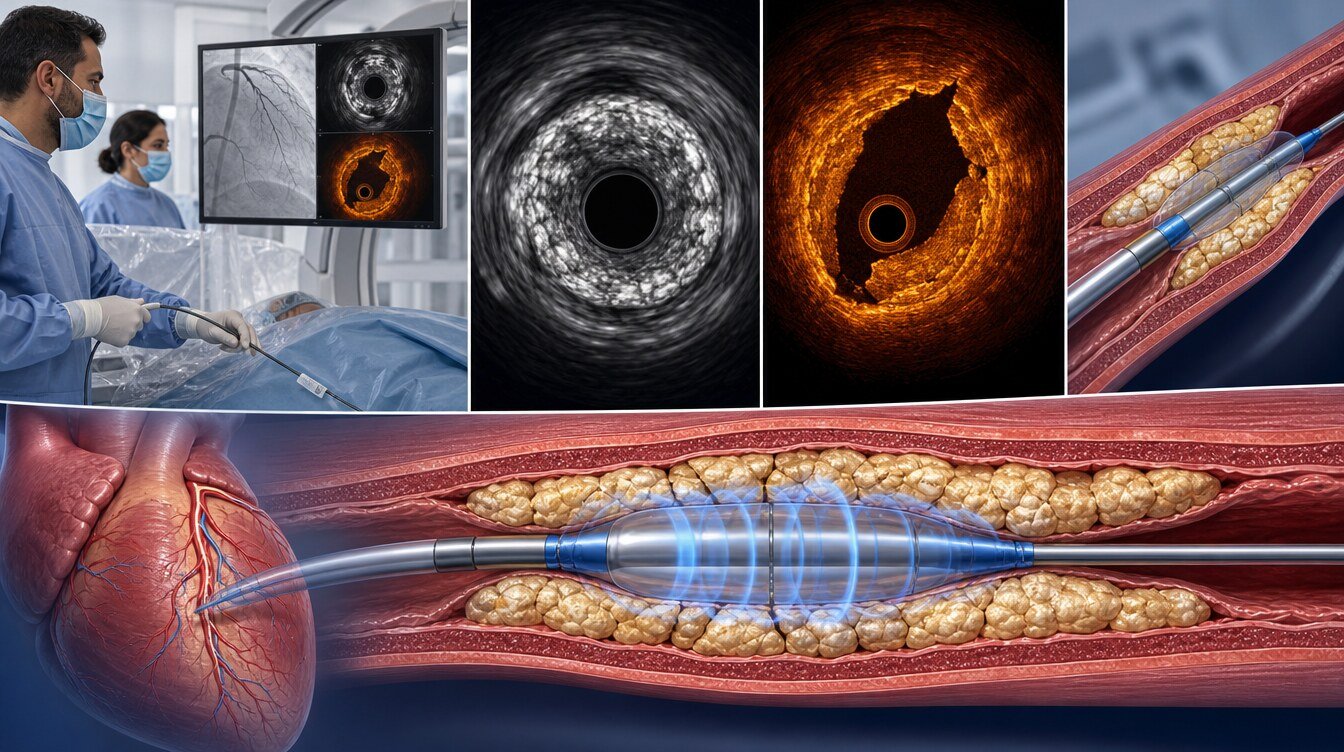
Coronary angiography gives a two-dimensional lumen view and can miss calcium depth, circumferential extent and true stent expansion. Intravascular ultrasound (IVUS) and optical coherence tomography (OCT) provide cross-sectional images that can help assess:[22,23]
- Whether calcium is superficial or deep, and how far it encircles the vessel
- Whether IVL, a balloon or a stent has created an adequate path or expansion
- Whether a calcium fracture is visible
- Whether the stent is apposed to the wall
Consensus texts discuss imaging as a guide to calcium phenotype and preparation choice. That does not mean imaging prevents heart attack or death, that it mandates IVL, or that it is technically possible in every patient. No universal expansion cut-off is published here.[22,23]
How is the procedure performed?
The procedure is done in a catheter laboratory equipped for coronary angiography and percutaneous coronary intervention (PCI). It is not open-heart surgery; the patient experience is similar to other coronary balloon–stent procedures:[27]
- Preparation: History, medicines, blood tests, kidney function and contrast risk are reviewed.
- Access: Radial or femoral artery under local anaesthesia.
- Imaging and wire: The artery is imaged; a fine wire is advanced beyond the stenosis.
- Preparation: The team chooses IVL, a balloon, rotational atherectomy, a stent or another tool according to the lesion and the live result. The IVL balloon is removed at the end.
- Result check: Flow, residual stenosis and, when used, IVUS/OCT findings are reassessed.
- Removal: Wires and catheters are withdrawn; the access site is controlled. If a stent was placed, the lasting structure is the stent.
Most patients remain awake. Brief chest discomfort during the procedure should be reported if new or severe.[27]
This page does not prescribe balloon diameter, pressure, pulse count, inflation cycles or device order.
What does the scientific evidence show?
Three ideas should be kept apart. Device or procedural success is whether the planned approach treats the lesion adequately. Calcium fracture, residual stenosis and minimum stent area are imaging results at the time of the procedure. Death, myocardial infarction and repeat procedures are separate, more patient-important endpoints. They are not interchangeable.[3,14]
The DISRUPT CAD family: single-arm foundational evidence
DISRUPT CAD I is a 60-person prospective feasibility cohort; it is not a randomised comparison. It is counted once within the family.[1]
DISRUPT CAD II is a 120-person multicentre single-arm post-approval study. IVL delivery/use was reported as 100% in the cohort; no abrupt closure, slow/no-reflow or perforation was observed during the procedure. Calcium fracture was seen in 78.7% of OCT-evaluated lesions. The primary endpoint is in-hospital MACE; all events were reported as non-Q-wave myocardial infarction (5.8%). It is single-arm and is not evidence of superiority over alternatives.[2]
DISRUPT CAD III is a single-arm regulatory study of 431 enrolled patients against prespecified performance goals. There is no concurrent randomised comparator arm. Procedural success was 92.4%; fracture was seen in 67.4% of OCT lesions; minimum stent area was 6.5±2.1 mm²; endpoints exceeded the performance goals. That is not superiority versus rotational atherectomy or balloons for death or myocardial infarction.[3]
The CAD III one-year report is follow-up of the same family (analysis n=384). MACE was 13.8%, cardiac death 1.1%, myocardial infarction 10.5%, ischaemia-driven target-lesion revascularisation 4.3%, and definite/probable stent thrombosis 1.1%. These are single-arm cohort observations, not comparative effects.[4]
DISRUPT CAD IV is a prospective regulatory study in Japan with 64 ITT patients. The comparison is a propensity-matched historical IVL control; it is not concurrent randomisation. Procedural success was 93.8%; perforation, abrupt closure or slow/no-reflow during the procedure was reported as 0.[5]
The CAD IV one-year paper is the same 64-person cohort. Cardiac death 0%, myocardial infarction 6.3%, target-vessel revascularisation 4.7% and stent thrombosis 0 were reported; there is no comparator arm.[6] The two-year paper is again the same cohort (62 people completed two years); MACE 12.6%, target-lesion revascularisation 3.2% and stent thrombosis 0 were reported. A DOI/PMID was not independently extracted in this package.[7]
The patient-level pooled CAD I–IV analysis includes 628 people. Thirty-day safety was 92.7% and effectiveness 92.4%; perforation, abrupt closure or no-reflow attributed to IVL was 0; serious angiographic complications were 2.1%. This analysis includes CAD I–IV patients; it is not added on top of those totals and has no comparator arm.[8]
The CAD I–II eccentric-calcium pooling includes 180 CAD I–II patients; it is not added to CAD I, CAD II or pooled I–IV totals.[9] The CAD III IVUS substudy is a secondary imaging record of the same CAD III cohort; numerical results were not extracted in this package and it is not counted as an independent trial.[10]
Randomised comparisons
ROTA.shock is a randomised comparison of rotational atherectomy with coronary IVL. This package has two separate publication identities (CCI and Am J Cardiol); whether the cohorts are the same was not verified. The accessed abstract did not provide an adequate supervised numerical endpoint, so no numerical “which is better” claim is made here.[11,12]
ROLLER COASTR-EPIC22 assigned 171 patients to rotational atherectomy, IVL or excimer laser (57 per arm). The primary endpoint is OCT percentage stent expansion. Minimum stent area was 5.4±1.8 mm² with IVL, 5.5±2.1 mm² with rotational atherectomy and 5.1±1.8 mm² with laser; IVL was noninferior to rotational atherectomy. No significant arm difference in procedural success or complications was reported. This is imaging/procedural noninferiority, not a mortality trial.[13]
BALI randomised 200 patients to conventional preparation with versus without added IVL (99/101). The one-year composite was 35% versus 52%; the authors note that the difference came mainly from the OCT residual area stenosis ≥20% component (32% versus 45%). It should not be interpreted as an isolated reduction in death, myocardial infarction or repeat procedures.[14]
BASIL compared IVL pretreatment with conventional balloon angioplasty in 60 patients. Procedural success was 72.7% versus 55.6% (P=.165); in-hospital MACE-free course was 90.9% versus 96.3% (P=.405), without a statistically significant difference. It is a small trial; identifiers were not independently verified in this package.[15]
EXIT-CALC is a 40-person single-centre RCT (19 IVL / 21 conventional). There was no significant difference in OCT minimum stent expansion (MSA 6.6±1.5 versus 6.2±1.8 mm²; P=.406); no MACE was reported at 30 days. “Zero events = no risk” must not be inferred from a very small sample. Identifiers are NA in this package.[16]
ICARE-OFDI is a 2026 randomised report comparing IVL with rotational atherectomy. The abstract reports a noninferior minimum stent area and equivalent 12-month target-lesion failure (TLF 2.4% IVL, 1.2% rotational atherectomy); full sample size and numerical MSA estimates are left NA in this package. It is not evidence of superiority for death or myocardial infarction.[17]
Registered, unpublished or ongoing studies
ISAR-CALC 2 is designed to compare a super high-pressure balloon with IVL after failed conventional NC preparation. Enrolment appears complete; this package has no verified results table, so clinical outcomes are not written.[19]
VICTORY is a registered comparison of IVL with OPN NC for OCT stent expansion in resistant lesions. Results had not been published at the cut-off date.[20]
The ShockFast study plans to compare the ShockFast device with Shockwave IVL on post-procedure OCT stent area. At the cut-off date the record is recruiting; there are no comparative results.[21]
2026 observational comparison
A TriNetX propensity-matched cohort compared IVL with atherectomy in single-vessel DES-PCI (13,499 before matching; 5,768 per arm). An association favouring IVL was reported for one-year all-cause death, myocardial infarction and MACE and for 30-day complications; the authors call for randomised confirmation. Residual confounding, selection, coding and unmeasured lesion complexity mean these estimates are not causal superiority or a personal risk reduction.[18]
What are possible benefits and limitations?
| Possible benefit / purpose | Limitation / required caveat |
|---|---|
| Calcium modification and stent preparation in a selected calcified stenosis | Does not remove calcium from the body; fracture or the same result is not guaranteed in every lesion |
| High procedural success and imaged fractures in DISRUPT CAD | Single-arm / performance-goal design; death/MI reduction was not shown |
| OCT-expansion noninferiority versus rotational atherectomy in ROLLER | An imaging endpoint; not a universal “which is better” ranking |
| Lower one-year composite in BALI | The difference comes mainly from the OCT residual-stenosis component |
| Balloon and catheter are removed at the end | The balloon must still cross; most de novo plans still consider a stent |
What are the risks?
IVL is used within coronary intervention. In addition to the general risks of balloon–stent procedures, complications specific to calcium modification are described. Some may relate to the device, some to the lesion and some to PCI in general:[8,27]
- Coronary dissection
- Coronary perforation
- Balloon rupture
- Slow flow or no-reflow
- Acute vessel closure
- Side-branch loss
- Transient cardiac-rhythm capture (the team monitors this)
- Myocardial infarction, stent thrombosis or arrhythmia
- Access-site bleeding and vascular complications
- Contrast-related kidney injury or allergy
- Later restenosis if a stent is used
The low angiographic-complication counts in DISRUPT CAD II and the pooled I–IV analysis are cohort observations; they are not a personal risk forecast or proof that “IVL is risk-free”.[2,8]
Transient capture of the heart rhythm can occur during the procedure; the team watches this on the monitor. That observation does not mean the procedure is risk-free for you.
What are the alternatives and complementary methods?
| Method | Core approach | Key difference from coronary IVL |
|---|---|---|
| Standard NC balloon | Controlled expansion without surface elements | Does not use acoustic waves |
| Scoring / cutting balloon | Focused preparation via surface elements or microblades | A separate balloon class; not mixed with IVL evidence |
| Super high-pressure NC | Selected resistant lesions not opened by a standard NC balloon | ISAR-CALC 2 / VICTORY results are unpublished in this package |
| Rotational atherectomy | Abrasion / path preparation with a diamond-coated burr | A different mechanism, independent of balloon crossing |
| Orbital atherectomy | A different atherectomy class | Must not be transferred to coronary IVL evidence |
| Excimer laser (ELCA) | Laser ablation | A separate arm in ROLLER; not equated with IVL |
| Peripheral IVL | Leg/peripheral vascular bed | Outside coronary IVL evidence |
| Kidney-stone lithotripsy | Extracorporeal shock waves | Not an intravascular coronary catheter procedure |
Rotational atherectomy and IVL may be combined in the same procedure (for example lesions in which both crossing and deep calcium are problems). Evidence for combined use is mainly registry/case-series level; it is not a routine prescription or proven superiority for every patient. Technical sequence is not described here.[22,23]
Preparation, discharge and recovery
Preparation depends on elective versus urgent setting, kidney function, bleeding risk, medicines, contrast history and access route. Follow hospital fasting and medicine instructions; do not stop prescribed medicines on your own.[27]
After planned uncomplicated coronary angioplasty some patients go home the same or next day. Local policy, clinical status and complications change the timing. This general frame comes from official PCI discharge patient information and is not a fixed discharge rule for every hospital in Türkiye.[27]
Discharge advice should be personalised and include access-site care, activity limits, antiplatelet and other prescriptions, follow-up, and when to seek emergency care.
Antiplatelet duration is set by the overall PCI, stent or drug-coated balloon strategy and the clinical picture, not by IVL alone. Stopping these medicines without advice can cause serious stent thrombosis.[24,27]
Bruising at the access site can occur. Increasing pain, swelling, redness, discharge or fever should prompt contact with the treating centre or another healthcare service.[27]
When should emergency services be called?
Call emergency services (112 in Türkiye) without delay for:[27]
- Chest pain that does not settle or is worsening
- Access-site bleeding that continues despite direct pressure or starts again
- Rapidly enlarging swelling at the access site
- Marked coldness, colour change or numbness in the treated arm or leg
- Fainting, severe breathlessness, sudden weakness or a change in consciousness
Not every bruise or mild tenderness is an emergency; uncontrolled bleeding, severe chest pain or impaired limb circulation is. Do not drive yourself with emergency symptoms. The 112 direction is a general safety frame; detailed local discharge wording should be reviewed against an official Turkish source before publication.[27]
Frequently asked questions
No. The one-year composite was lower with IVL; the difference came mainly from the OCT residual area stenosis component. It is not an isolated reduction in clinical events.[14]
Assessment and decision
Coronary IVL is a temporary preparation tool used in selected calcified stenoses. Planning is broader than “will IVL be used?” and includes treatment need, balloon crossing, calcium morphology, the stent plan, IVUS/OCT findings, alternatives and centre experience.[22–25]
The 2021 ACC/AHA/SCAI and 2024 ESC documents have no IVL-specific Class/LOE recommendation verified in this package (NA). EAPCI and SCAI texts are consensus statements and must not be presented as graded guideline recommendations. The decision rests on individual clinical assessment.[22–25]
Literature
References
Brinton TJ, et al. Feasibility of Shockwave Coronary Intravascular Lithotripsy for the Treatment of Calcified Coronary Stenoses. Circulation. 2019. DOI: 10.1161/CIRCULATIONAHA.118.036531. PMID: 30715944. Trial: NCT02923193.
Ali ZA, et al. Safety and Effectiveness of Coronary Intravascular Lithotripsy for Treatment of Severely Calcified Coronary Stenoses: The Disrupt CAD II Study. Circ Cardiovasc Interv. 2019. DOI: 10.1161/CIRCINTERVENTIONS.119.008434. Trial: NCT03328949. A PMID was not independently resolved in this package.
Hill JM, et al. Intravascular Lithotripsy for Treatment of Severely Calcified Coronary Artery Disease: The Disrupt CAD III Study. J Am Coll Cardiol. 2020. DOI: 10.1016/j.jacc.2020.09.603. PMID: 33069849. Trial: NCT03595176.
Kereiakes DJ, et al. Intravascular Lithotripsy for Treatment of Severely Calcified Coronary Lesions: 1-Year Results From the Disrupt CAD III Study. JSCAI. 2022. DOI: 10.1016/j.jscai.2021.100001. PMID: 39130140. PMCID: PMC11308114. Trial: NCT03595176. Follow-up of the same CAD III cohort.
Saito S, et al. Intravascular Lithotripsy for Vessel Preparation in Severely Calcified Coronary Arteries Prior to Stent Placement: Primary Outcomes From the Japanese Disrupt CAD IV Study. Circ J. 2021. DOI: 10.1253/circj.CJ-20-1174. PMID: 33551398. Trial: NCT04151628.
Saito S, et al. Japanese Disrupt CAD IV Study 1-Year Results. Circ Rep. 2022. DOI: 10.1253/circrep.CR-22-0068. PMID: 36120480. PMCID: PMC9437473. Trial: NCT04151628. The same CAD IV cohort.
Saito S, et al. Japanese Disrupt CAD IV Study 2-Year Results. Circ Rep. 2023. Trial: NCT04151628. A DOI/PMID was not independently extracted in this package. The same CAD IV cohort.
Kereiakes DJ, et al. Intravascular Lithotripsy for Treatment of Calcified Coronary Lesions: Patient-Level Pooled Analysis of the Disrupt CAD Studies. Cardiovasc Revasc Med. 2021. Includes CAD I–IV patients; not an independent cohort. A DOI/PMID was not verified in this package.
Blachutzik F, et al. Safety and effectiveness of coronary intravascular lithotripsy in eccentric calcified coronary lesions: a patient-level pooled analysis from the Disrupt CAD I and CAD II Studies. Clin Res Cardiol. 2020. DOI: 10.1007/s00392-020-01737-3. PMID: 32948882. PMCID: PMC7862504. Includes CAD I–II patients; not added to the totals.
DISRUPT CAD III IVUS substudy. Cardiovasc Revasc Med. 2023. DOI: 10.1016/j.carrev.2023.03.003. PMID: 36934007. Secondary imaging record of the same CAD III cohort; numerical results were not extracted in this package.
Blachutzik F, et al. Coronary intravascular lithotripsy and rotational atherectomy for severely calcified stenosis: Results from the ROTA.shock trial. Catheter Cardiovasc Interv. 2023. DOI: 10.1002/ccd.30815. PMID: 37668088. No numerical comparative claim is made in this package.
Blachutzik F, et al. Comparison of Coronary Intravascular Lithotripsy and Rotational Atherectomy in the Modification of Severely Calcified Stenoses. Am J Cardiol. 2023. DOI: 10.1016/j.amjcard.2023.02.028. PMID: 37012181. Cohort overlap with the CCI report was not verified in this package; the reports are not pooled.
Jurado-Román A, et al. Rotational Atherectomy, Lithotripsy, or Laser for Calcified Coronary Stenosis: The ROLLER COASTR-EPIC22 Trial. JACC Cardiovasc Interv. 2025. DOI: 10.1016/j.jcin.2024.11.012. PMID: 39918495. Trial: NCT04181268.
Kristensen AT, et al. Balloon Lithotripsy Added to Conventional Preparation Before Stent Implantation in Severely Calcified Coronary Lesions. JACC Cardiovasc Interv. 2025. DOI: 10.1016/j.jcin.2025.09.028. Trial: NCT04253171. A PMID was not verified in this package.
Wong B, et al. Balloon angioplasty versus Shockwave intravascular lithotripsy in calcified coronary arteries: the BASIL study. J Invasive Cardiol. 2025. DOI/PMID/URL were not independently verified in this package.
Oomens T, et al. EXpansion of stents after intravascular lithoTripsy versus conventional predilatation in CALCified coronary arteries (EXIT-CALC). 2023. DOI/PMID/URL were not verified in this package.
Honton B, et al. Intravascular lithotripsy in comparison to rotational atherectomy for calcified lesions: the ICARE OFDI randomised trial. EuroIntervention. 2026. PMID: 42200665. Sample size and full MSA figures are NA in this package.
Miks C, Ozaki G, Shukla CR, Sharma V. Intravascular Lithotripsy Is Associated With Superior Clinical Outcomes Compared to Atherectomy: A Large-Scale, Propensity-Matched Analysis. Catheter Cardiovasc Interv. 2026. DOI: 10.1002/ccd.70464. PMID: 41837695. Observational association; not causal superiority.
ClinicalTrials.gov. ComparIson of Strategies to PrepAre SeveRely CALCified Coronary Lesions 2 (ISAR-CALC 2). NCT05072730. No verified results table.
ClinicalTrials.gov. The Value of IVL Compared To OPN Non-Compliant Balloons for Treatment of RefractorY Coronary Lesions (VICTORY). NCT05346068. Results have not been published.
ClinicalTrials.gov. ShockFast Intravascular Lithotripsy Device for Treatment of Calcified Coronary Lesions. NCT07407738. Recruiting; no comparative results.
Barbato E, et al. Management strategies for heavily calcified coronary stenoses: an EAPCI clinical consensus statement in collaboration with the EURO4C-PCR group. Eur Heart J. 2023. DOI: 10.1093/eurheartj/ehad342. PMID: 37208199. No IVL-specific Class/LOE is transferred from this package.
Riley RF, et al. SCAI Expert Consensus Statement on the Management of Calcified Coronary Lesions. JSCAI. 2024. DOI: 10.1016/j.jscai.2023.101259. PMID: 39132214. PMCID: PMC11307856. No IVL-specific Class/LOE was verified in this package.
Lawton JS, et al. 2021 ACC/AHA/SCAI Guideline for Coronary Artery Revascularization. J Am Coll Cardiol. 2022. DOI: 10.1016/j.jacc.2021.09.006. No IVL-specific Class/LOE was verified in this package (NA).
Vrints C, et al. 2024 ESC Guidelines for the management of chronic coronary syndromes. Eur Heart J. 2024. DOI: 10.1093/eurheartj/ehae177. PMID: 39210710. No IVL-specific Class/LOE was verified in this package (NA).
Shockwave Medical. Shockwave C2 Coronary IVL System Instructions for Use. Device identity / instructions for use. Not used as clinical-superiority evidence.
MedlinePlus. Angioplasty and stent - heart - discharge. Official patient information. Accessed 27 August 2026.
Continue