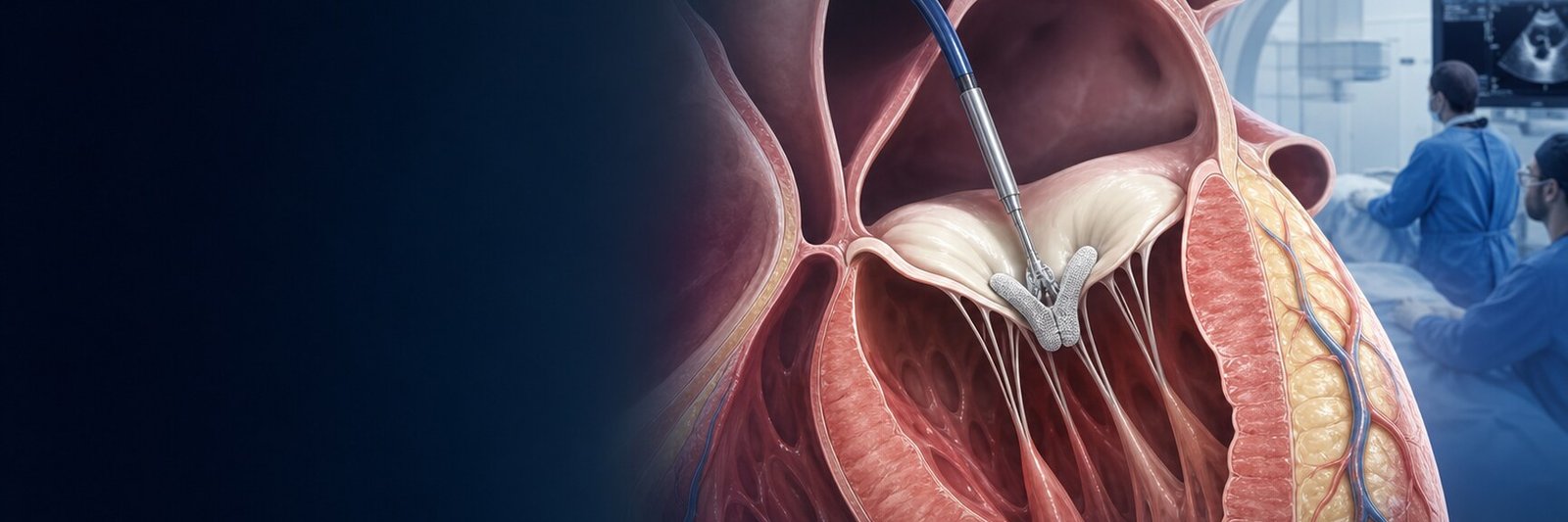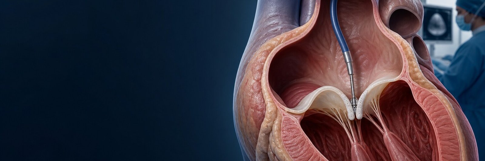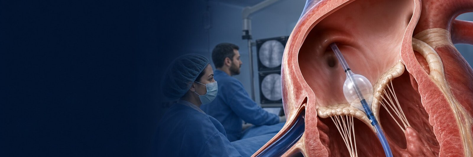
Mitral balloon valvuloplasty is a catheter procedure intended to separate fused commissures in suitable rheumatic mitral stenosis. Guidelines often use percutaneous mitral commissurotomy (PMC); PMBC, PTMC and PBMV may describe the same general intervention.[1,2]
What is mitral stenosis?
Mitral stenosis obstructs flow from the left atrium to the left ventricle, potentially causing breathlessness, atrial fibrillation, pulmonary hypertension and embolic risk. Rheumatic commissural fusion differs from degenerative mitral-annular-calcification stenosis; balloon commissurotomy is not routinely generalised to the latter.[1]
How are severity and symptoms interpreted together?
Valve area, mean gradient and pulmonary pressure are affected by heart rate, rhythm and flow. No single number is interpreted alone. When symptoms and resting echocardiography disagree, exercise testing or exercise echocardiography can assess symptoms, gradient and pulmonary pressure under stress.[1,2]
Who may be assessed?
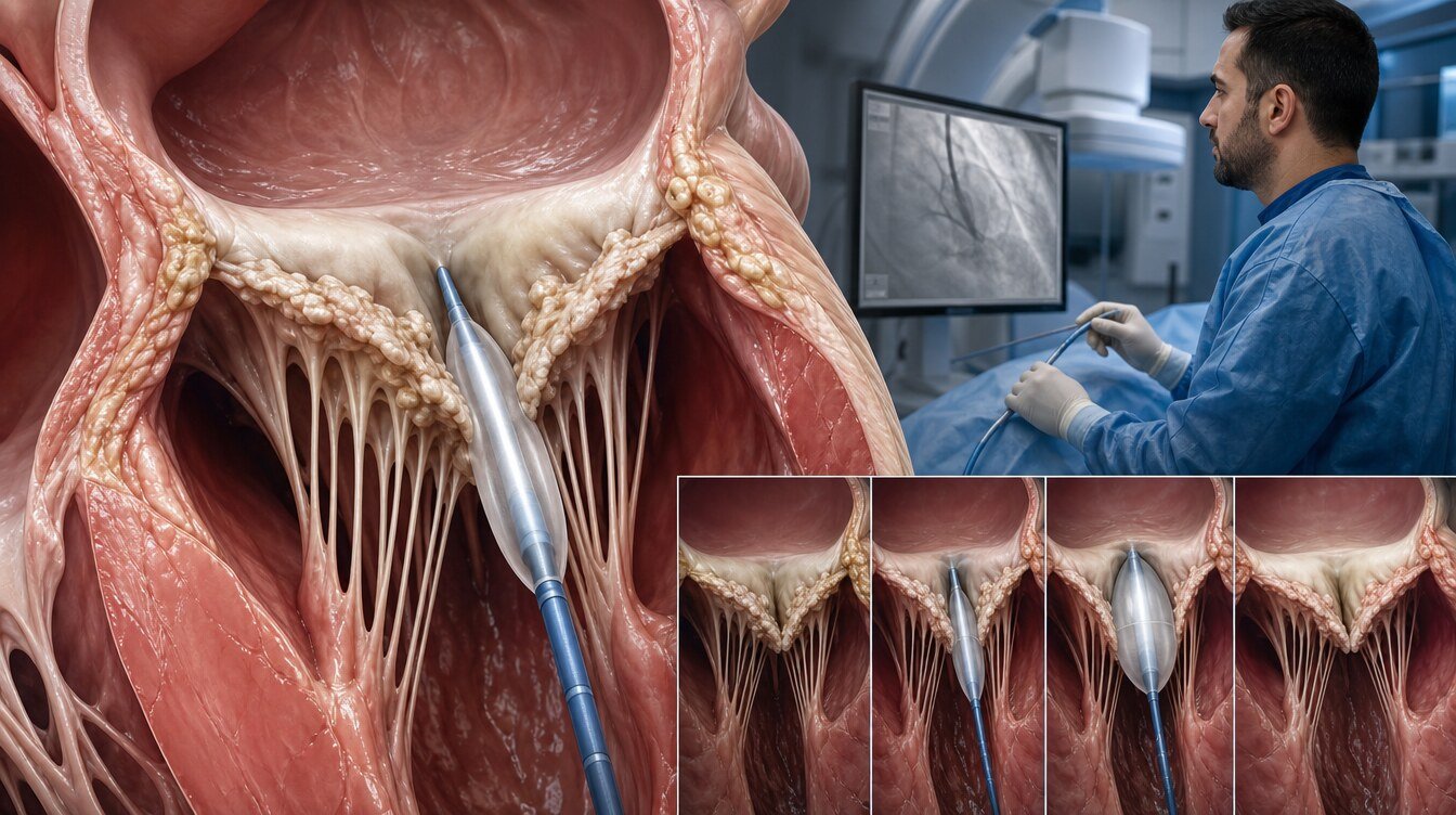
PMC is an established option for symptomatic clinically severe rheumatic mitral stenosis with suitable anatomy. Selected asymptomatic patients with high thromboembolic or haemodynamic risk may also be assessed.[1]
Major contraindications include left-atrial thrombus, more than mild MR, severe or bicommissural calcification, absent commissural fusion, severe subvalvular disease and another valve or coronary disease requiring surgery.[1]
The Wilkins score is not the whole decision. Balloon treatment works by separating fused commissures; absent fusion leaves no appropriate target. Commissural calcium, leaflet mobility and calcification, subvalvular disease, baseline MR and left-atrial thrombus are assessed together.[1,3]
| Favourable feature | Feature that may limit benefit or safety |
|---|---|
| Clear commissural fusion | No commissural fusion |
| Mobile, minimally calcified leaflets | Severe or bicommissural calcium |
| Limited subvalvular disease | Advanced chordal/papillary involvement |
| No more than mild MR | More than mild MR |
| No left-atrial thrombus | Left-atrial/appendage thrombus |
Balloon treatment or surgery?
Suitable rheumatic anatomy may be treated without sternotomy and may defer prosthetic valve replacement. Surgery may be favoured with important MR, heavy calcification, absent fusion, coronary bypass needs or another valve operation. Open commissurotomy and valve replacement are different surgical strategies; prosthesis choice adds separate lifetime considerations.[1,2]
How is the procedure performed?
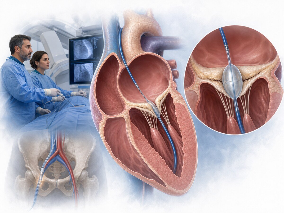
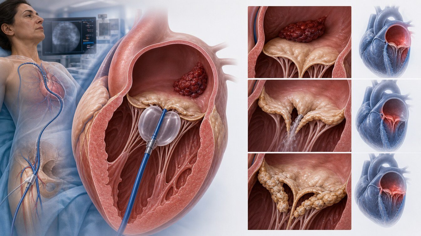
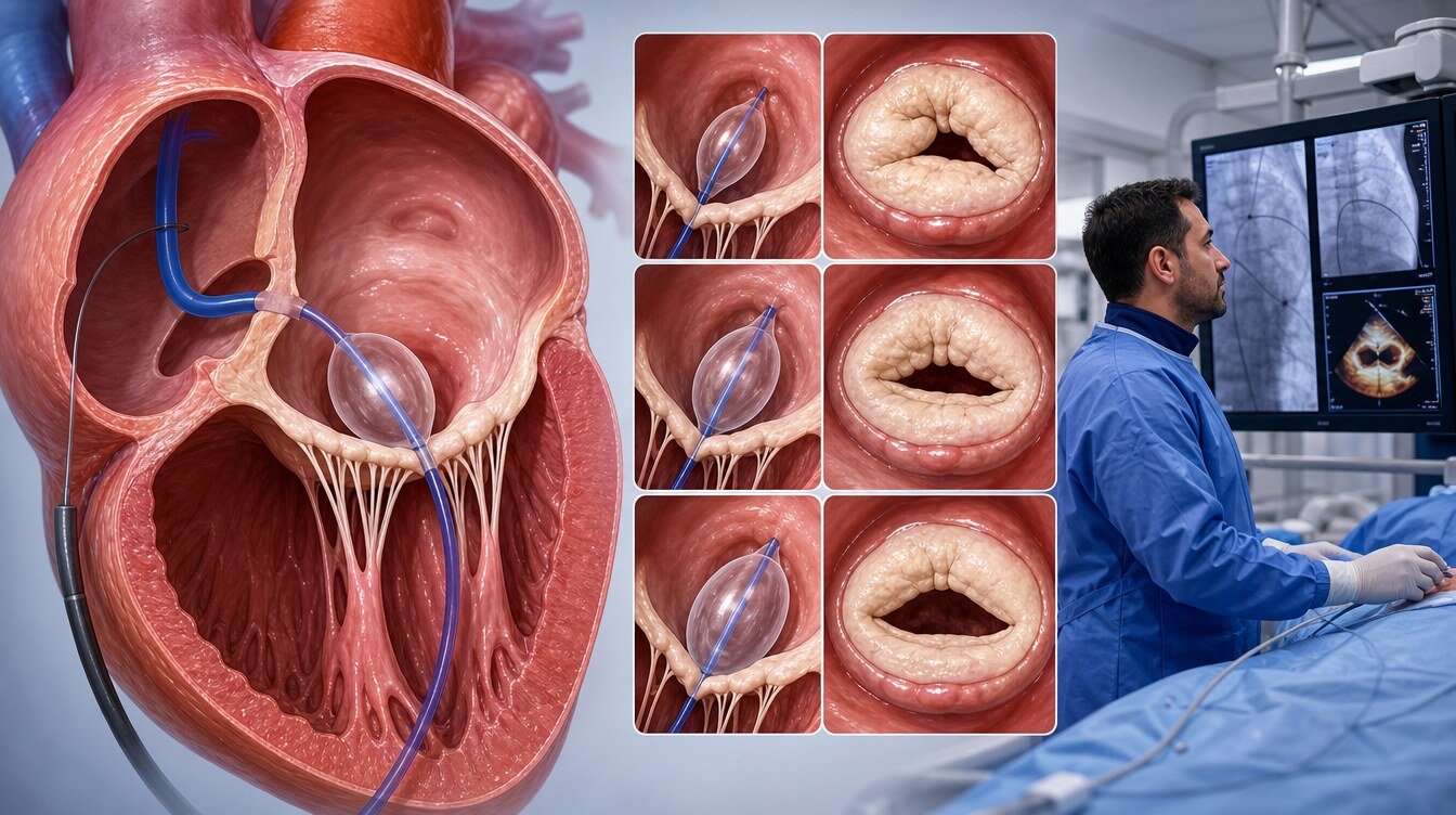
Venous access is obtained through the groin, followed by controlled transseptal passage. A dedicated balloon is advanced across the mitral valve and inflated in stages to separate fused commissures. Valve area, gradient and MR are reassessed before the catheter is removed.
Balloon sizing and staged inflation are adapted to body size, anatomy and intraprocedural findings. The aim is not maximal opening at any cost but a controlled balance between improved valve opening and the risk of new MR.
Benefits, risks and follow-up
Successful treatment can increase valve area, reduce gradient and improve symptoms. Rheumatic disease is not eliminated; restenosis and later repeat balloon treatment or surgery may occur.[3–6]
Procedural success combines commissural opening, greater valve area, lower gradient and atrial pressure without important new MR or a major complication. Clinical success also considers breathlessness and functional capacity. Durability varies with anatomy, post-procedure valve area and MR, atrial fibrillation, pulmonary hypertension and progressive calcification.[3–6]
Pregnancy planning and atrial fibrillation
The increased flow and heart rate of pregnancy can unmask or worsen mitral stenosis. In clinically severe rheumatic disease, pre-pregnancy assessment is important; intervention during pregnancy requires an experienced Pregnancy Heart Team and careful maternal, fetal and radiation planning.[1]
Atrial fibrillation increases thrombus and embolic risk. Balloon treatment does not automatically restore rhythm or remove the need for anticoagulation; these decisions remain separate.[1,2]
Risks include severe MR and urgent surgery, tamponade, stroke or systemic embolism, access-site bleeding, residual atrial septal shunt, arrhythmia, inadequate opening, restenosis, infection, kidney injury and death.
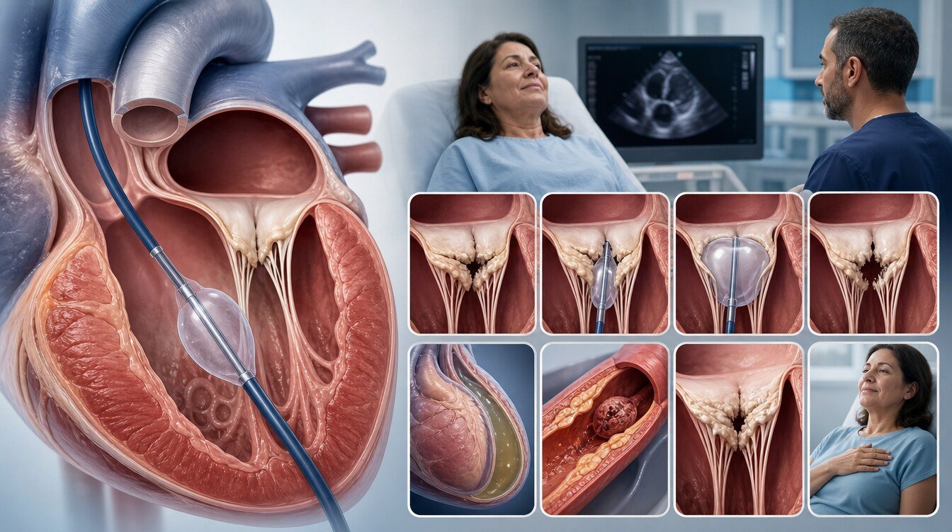
After the procedure, rhythm, access site, MR and pericardial findings are monitored. Anticoagulation and rhythm management are individual. Symptoms and echocardiography remain under follow-up.
Follow-up also evaluates pulmonary pressure, right-heart response and recurrent stenosis. If restenosis occurs, commissural anatomy and MR are reassessed before repeat balloon treatment or surgery is considered.
Questions to ask the Heart Team
- Is the stenosis rheumatic commissural fusion or degenerative calcification?
- How do valve area, gradient and pulmonary pressure fit together?
- Is there left-atrial thrombus or more than mild MR?
- Which commissural or calcification finding matters beyond the Wilkins score?
- How will balloon size and the stopping point be selected?
- What is the emergency plan if severe MR develops?
- If restenosis develops, would repeat balloon treatment or surgery be considered?
Frequently asked questions
No. It is a transseptal catheter-based percutaneous mitral commissurotomy.
No. The principal setting is rheumatic stenosis with commissural fusion; it is not routinely generalised to degenerative calcific stenosis.[1]
Left-atrial thrombus is a major contraindication; treatment and reassessment are individual.[1]
More than mild MR is generally a contraindication because the procedure may worsen regurgitation.[1]
It can enlarge the valve opening and reduce symptoms but does not remove rheumatic disease; restenosis can occur.
The plan depends on atrial fibrillation, prior thrombus or embolism and other indications.
It may be considered in selected severely symptomatic patients despite medical therapy at an expert centre with a Pregnancy Heart Team.[1]
Timing depends on access-site, rhythm and echocardiographic findings; follow the individual discharge plan.
Assessment and decision
Balloon commissurotomy can be effective in the right rheumatic anatomy; “balloon for every mitral stenosis” is incorrect.
Literature
References
Praz F, et al. 2025 ESC/EACTS Guidelines. doi:DOI: 10.1093/eurheartj/ehaf194.
Otto CM, et al. 2020 ACC/AHA Guideline. doi:DOI: 10.1161/CIR.0000000000000923.
Bouleti C, et al. Calcification and long-term PMC results. doi:DOI: 10.1161/CIRCINTERVENTIONS.113.000858.
Reyes VP, et al. Balloon versus open commissurotomy. doi:DOI: 10.1056/NEJM199410133311501.
Meneguz-Moreno RA, et al. Very long-term PBMV follow-up. PMID 30077684.
Dadjo Y, et al. Mid-term PTMC outcomes. doi:DOI: 10.1186/s12872-021-02175-3.
This content is for general information only and does not replace personalized medical advice. In emergencies call local emergency services.
Continue