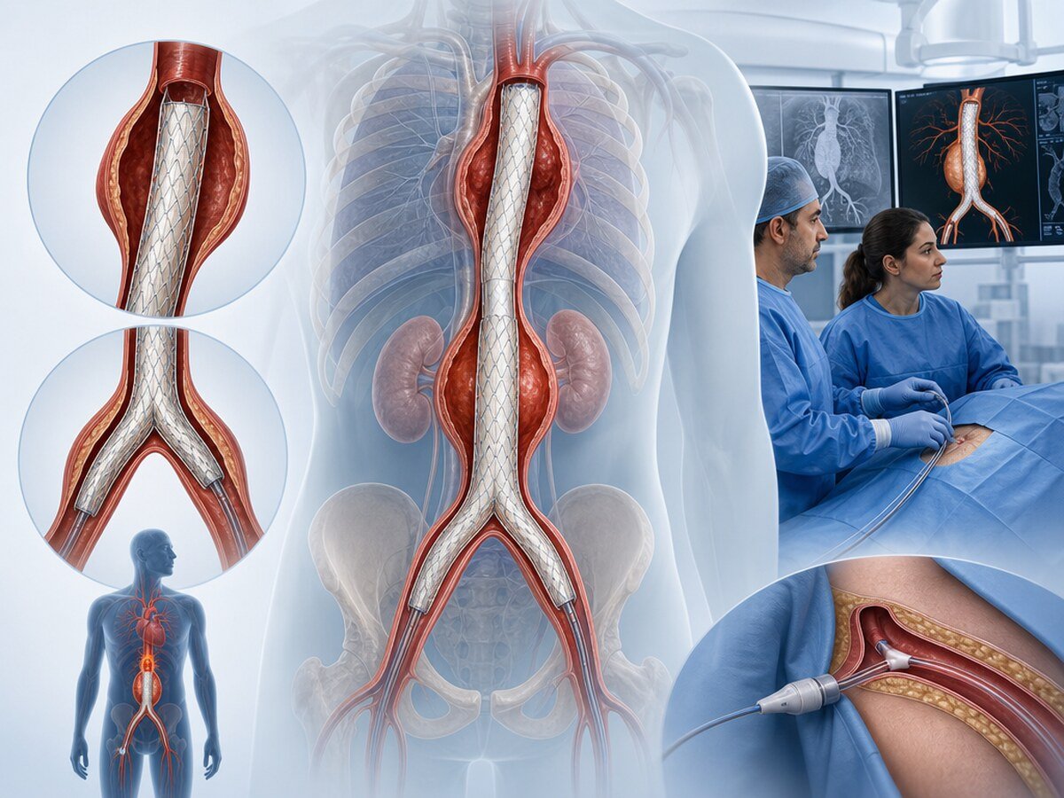
An aortic aneurysm is widening of the wall of the aorta, the main artery that leaves the heart and carries blood through the body, beyond its usual size. Endovascular treatment places a covered support called a stent-graft inside the aorta through a catheter so that the diseased segment is excluded from the blood flow. Not every aneurysm is the same; surveillance, open surgery or endovascular treatment is chosen according to the segment, shape, growth rate, symptoms, general health and vessel anatomy.[1,2]
Abdominal and thoracic aortic aneurysms can look as if they share a mechanism, but treatment thresholds, anatomy, rupture risk and the procedures that can be used differ. A diameter cut-off from the abdominal aorta is therefore not transferred to the thoracic aorta.[1–4]
This text is general patient education; it is not a diagnosis or personal treatment advice. It requires a final physician review before publication.
What is the aorta, and what does aneurysm mean?
The aorta is the largest artery. It leaves the left ventricle and travels through the chest and abdomen to supply the brain, arms, organs and legs. Its wall has three layers:
- Intima: the inner layer in contact with blood
- Media: the middle layer that gives strength and elasticity
- Adventitia: the outer connective-tissue layer
An aneurysm is a lasting widening or ballooning of part of the vessel wall. That widening can reduce mechanical strength. High blood-pressure strain, breakdown of structural proteins, inflammation, atherosclerosis or inherited connective-tissue disease can take part. Smoking, ageing and some genetic traits can make the process easier.[1,2]
An aneurysm often grows slowly and causes no symptoms for a long time. Marked widening, rapid growth, a tear inside the wall or bleeding can become life-threatening.
Abdominal and thoracic aortic aneurysm: two different disease groups
Abdominal aortic aneurysm
An abdominal aortic aneurysm (AAA) forms in the part of the aorta below the diaphragm. The infrarenal aorta, below the renal arteries, is most often involved. The aneurysm can also reach the origin of the iliac arteries.[2,3]
Most abdominal aortic aneurysms are found by chance on ultrasound, computed tomography or other imaging. Large or growing aneurysms can cause abdominal or back pain and, less often, a pulsatile mass around the navel.
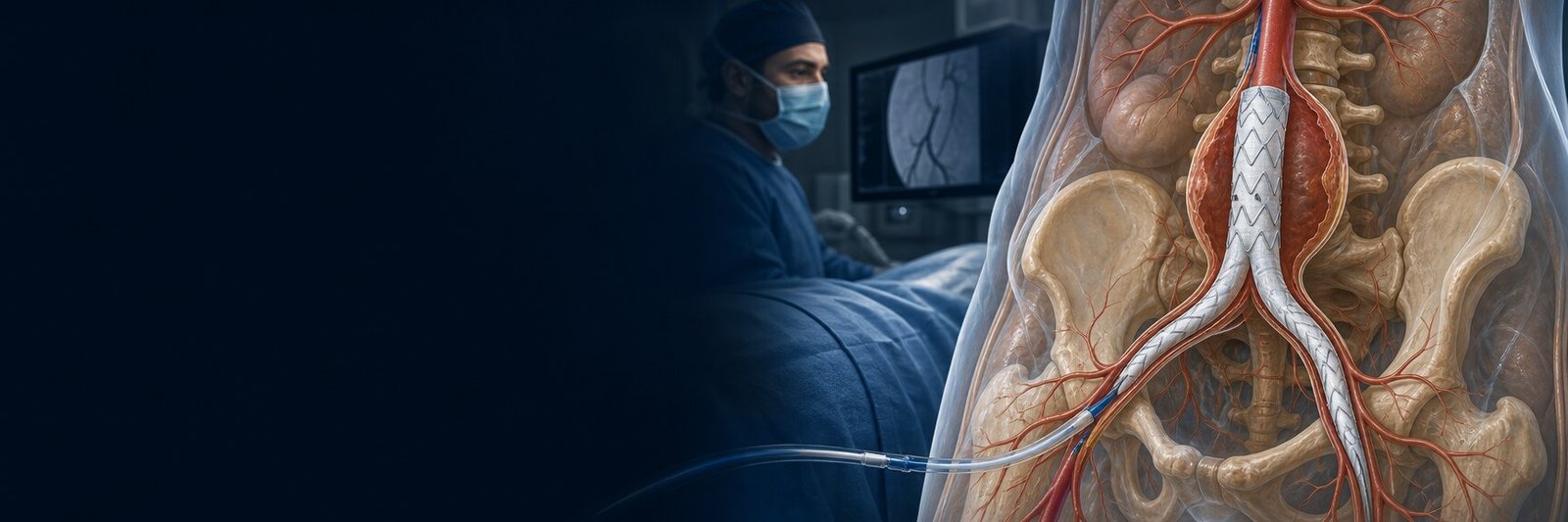
In suitable anatomy, endovascular repair of the abdominal aorta is considered as EVAR. Planning, endoleak types and randomised evidence belong on that page.
Thoracic aortic aneurysm
A thoracic aortic aneurysm (TAA) develops in the part of the aorta inside the chest. The root and ascending aorta, the arch or the descending thoracic aorta can be involved. Branch vessels, blood flow to the heart and brain, and surgical access differ in each region.[1,4]
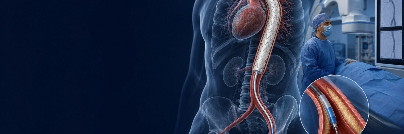
A thoracic aortic aneurysm can cause chest or back pain, hoarseness, difficulty swallowing or breathlessness; most cases are still silent. Endovascular treatment of the descending thoracic aorta is usually called TEVAR. TEVAR has separate evidence for dissection, ulcer, trauma or rupture; those results are not transferred to AAA.
This page is not the same picture as peripheral artery disease. Evidence from leg-artery narrowing is not transferred to aortic aneurysm.
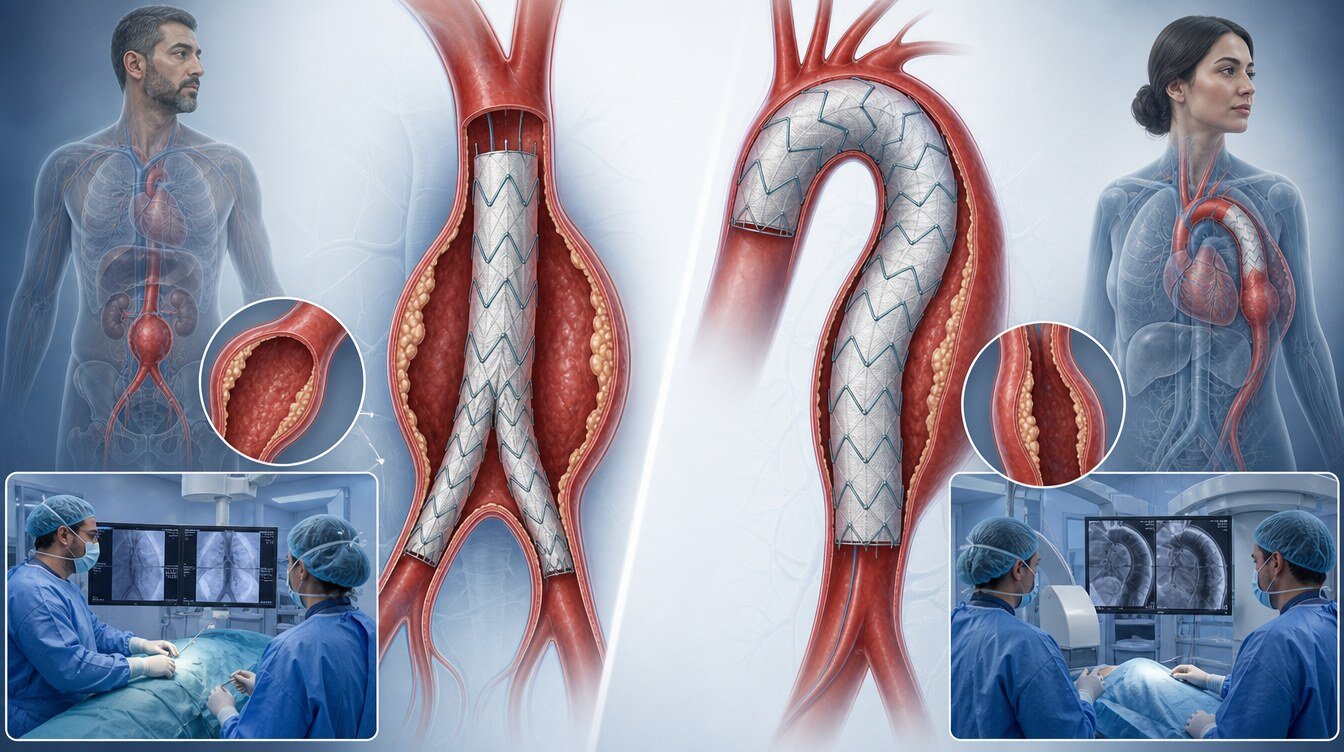
Different forms of aneurysm
A true aneurysm is ballooning in which all three layers of the aortic wall widen together. The classic spindle-shaped fusiform aneurysm belongs here. Saccular aneurysms that bulge as a pouch can also be true aneurysms; they can carry a different risk in some settings.
A false aneurysm or pseudoaneurysm is a “false” balloon after the wall has torn and blood is contained by surrounding tissue, clot or a surgical field. The three layers have not widened as an intact wall. It can follow earlier surgery, a catheter procedure, trauma, infection or a suture-line problem.
Dissection-related dilatation follows a tear in the inner layer and blood travelling between the wall layers, creating a false lumen. Treatment of dissection is assessed differently from an ordinary fusiform aneurysm.[1]
Rupture is complete tearing of the wall so that blood leaks into the body. Rupture of an abdominal aortic aneurysm usually causes rapid bleeding behind or into the abdomen and is a surgical emergency.
Diameter thresholds for these types are not interchangeable. Rupture risk is not set by diameter alone; shape, wall structure, growth rate, pain, dissection, infection, the aortic segment and body size also matter.[1–3]
Risk factors and inherited aortic disease
Features that can raise the risk of aortic aneurysm include:
- Older age
- Male sex
- Current or previous heavy smoking
- High blood pressure
- Atherosclerosis
- Aortic aneurysm or dissection in a first-degree relative
Stopping smoking, keeping blood pressure at the target set by a clinician and reducing atherosclerotic risk matter for both general cardiovascular health and aortic care.[1,2] Cholesterol (hyperlipidaemia) treatment does not “shrink” the aortic wall; it is part of the overall cardiovascular-risk plan.
When is a genetic assessment considered?
The following findings raise the chance of inherited thoracic aortic disease:[6]
- Family aortic dissection, aneurysm or unexplained sudden death
- Aneurysm or dissection at a young age
- Marfan syndrome
- Loeys–Dietz syndrome
- Vascular Ehlers–Danlos syndrome
- A bicuspid aortic valve
- Aortic disease in more than one vascular territory
Genetic counselling, suitable testing and screening of relatives may then be discussed. Family screening usually means cardiology review and suitable imaging of first-degree relatives.
Why do sex and body size matter?
The starting aortic diameter is on average smaller in women, so the same absolute diameter may not mean the same thing as in men. Some data suggest that rupture can occur at smaller diameters in women. Indexed assessment by body-surface area can therefore be more informative, especially in women.[2,3,7]
Symptoms and incidental detection
Most aneurysms cause no symptoms. They may be found when ultrasound, computed tomography or magnetic resonance imaging is done for another reason.
Possible symptoms include:
- Ongoing or new pain in the abdomen or loin
- A pulsatile mass around the navel
- Pain in the chest or back
- Hoarseness
- Difficulty swallowing
- Breathlessness or cough
These symptoms also occur in other diseases; new, unexplained or severe pain should not be delayed, especially in a person already known to have aortic disease.
Aortic imaging
Ultrasound uses sound waves and does not use radiation. It is often used to screen and follow abdominal aortic aneurysms. Guidelines recommend a one-time ultrasound screen in men aged 65–75 who have ever smoked; in other groups the decision follows individual risk.[2,3]
CT angiography (CTA) maps the arteries in detail, usually with iodinated contrast. It is fast in emergencies and important for stent-graft planning, branch vessels and access routes.
MR angiography (MRA) uses a magnetic field and radio waves and does not use radiation. It can be an alternative when repeated CT or contrast or radiation is a concern; it is not suitable for every person or every emergency.
How is diameter measured?
Aortic diameter is usually measured outer wall to outer wall. Measurement perpendicular to the centreline is preferred. On axial CT, the minor axis is often closer to the true maximum diameter than the longest axial diameter. When measurements from different centres or techniques are compared, the method should be kept the same.[2]
Surveillance intervals
The interval follows diameter, growth rate, segment, shape and accompanying disease. For small infrarenal AAA, guidelines give these example intervals:[2]
| Measured diameter | Usual surveillance approach |
|---|---|
| 2.6–2.9 cm | A review at about 10 years may be considered |
| 3.0–3.9 cm | About every 3 years |
| 4.0–4.9 cm | About once a year |
| 5.0–5.4 cm | About every 6 months |
These are not an automatic calendar for every person. Measurement uncertainty, rapid growth, a saccular shape, female sex, a family history or genetic disease can require closer review. Intervals for the thoracic aorta are separate.
Stable aneurysm versus an emergency aortic state
Elective assessment means the aneurysm is not causing symptoms, there is no sign of rupture, and a procedure can be prepared. The aim is to weigh rupture risk against procedural risk.
New pain, clear growth over a short time, a leak in the wall or suspected rupture need faster assessment. A leak is limited escape of blood from the wall into surrounding tissue; it can appear before complete rupture.
Treatment options
No method is universally superior for every person.[1–4]
Surveillance and cardiovascular-risk reduction. Small, stable, asymptomatic aneurysms are imaged at intervals. Smoking should stop completely, blood pressure should stay at the clinician’s target, a statin may be considered according to atherosclerotic risk, and diabetes and weight should be managed. These steps do not necessarily shrink the aneurysm; the aim is to reduce growth and overall cardiovascular events.[1,2]
Open surgical repair. The diseased aortic segment is replaced with a prosthetic graft through an abdominal or chest incision. It is a larger operation; recovery is longer and early heart, lung, kidney or other organ risks can be higher. In suitable people it can provide a durable repair.
EVAR. Endovascular abdominal aortic repair, usually from the femoral arteries in the groin. In selected people with suitable anatomy, randomised trials have generally shown a lower early peri-procedural complication and death risk than open repair, with overall survival becoming similar by about two years. Lifelong imaging and occasional reintervention may be needed after EVAR.[2,3,8] Detail is on the EVAR page.
TEVAR. Thoracic endovascular aortic repair, used especially for suitable lesions of the descending thoracic aorta. For that segment, TEVAR can have advantages in early hospital stay and some complications compared with open surgery. Evidence quality varies by disease type; genetic aortopathy, the root, the ascending aorta or extensive thoraco-abdominal disease may need a different approach.[1,4] Detail is on the TEVAR page.
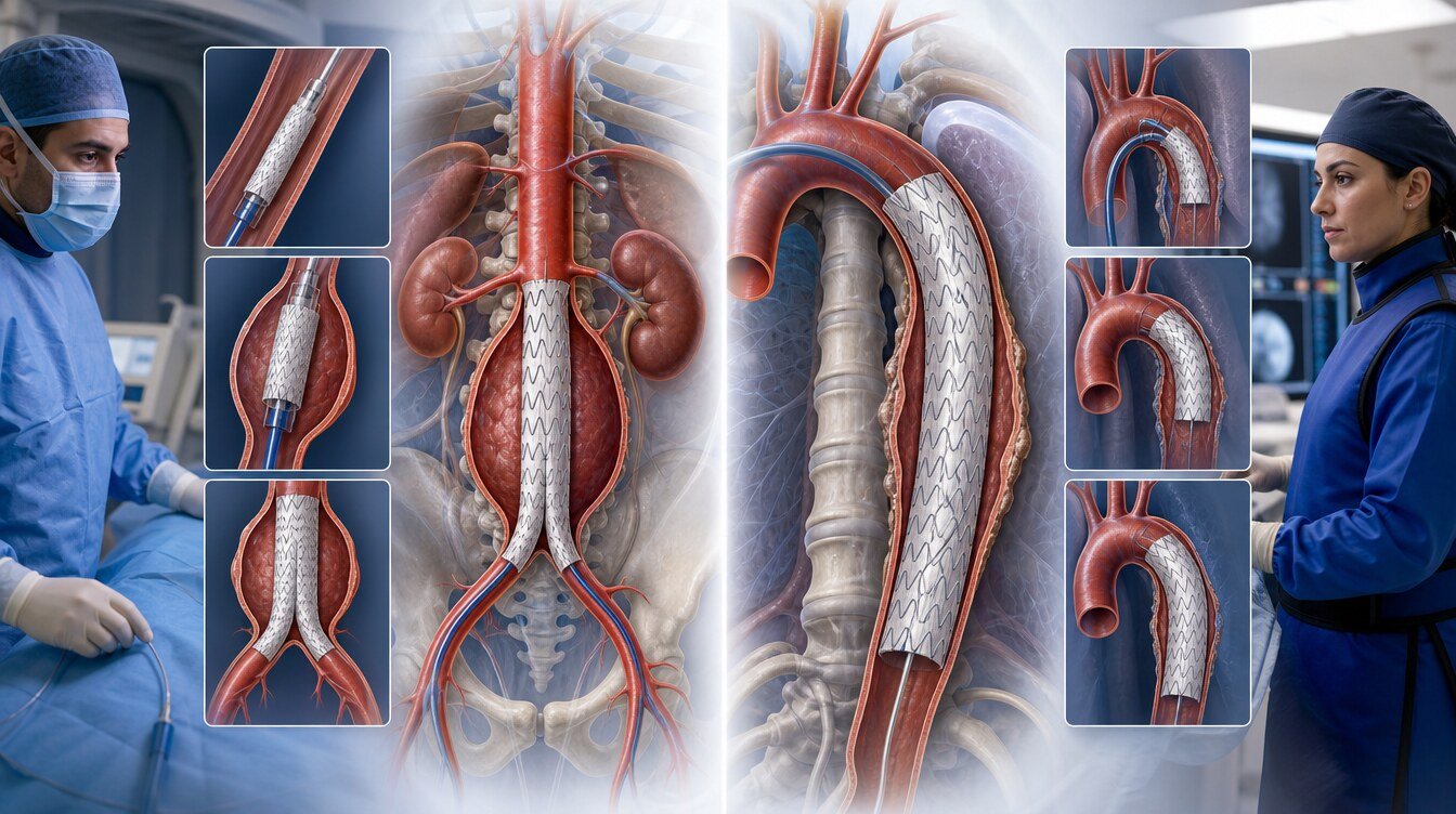
What is the treatment decision based on?
The decision brings these elements together:[1–4]
- Aortic segment and aneurysm length
- Maximum diameter and how it was measured
- Growth rate
- Pain, embolus, leak or rupture
- Whether the aneurysm is fusiform, saccular, a pseudoaneurysm or dissection-related
- Position of branch vessels
- Suitability of the femoral and iliac arteries for access
- Kidney, heart and lung function
- Age, life expectancy and daily function
- Genetic aortic disease
- Centre experience and a multidisciplinary aortic team
- The person’s values and preferences
For infrarenal fusiform AAA, guideline examples used when planned repair is discussed are about 5.5 cm in men and 5.0 cm in women. These are not universal or automatic operation limits; they change with segment, sex, body size, growth, symptoms and overall risk.[2,3]
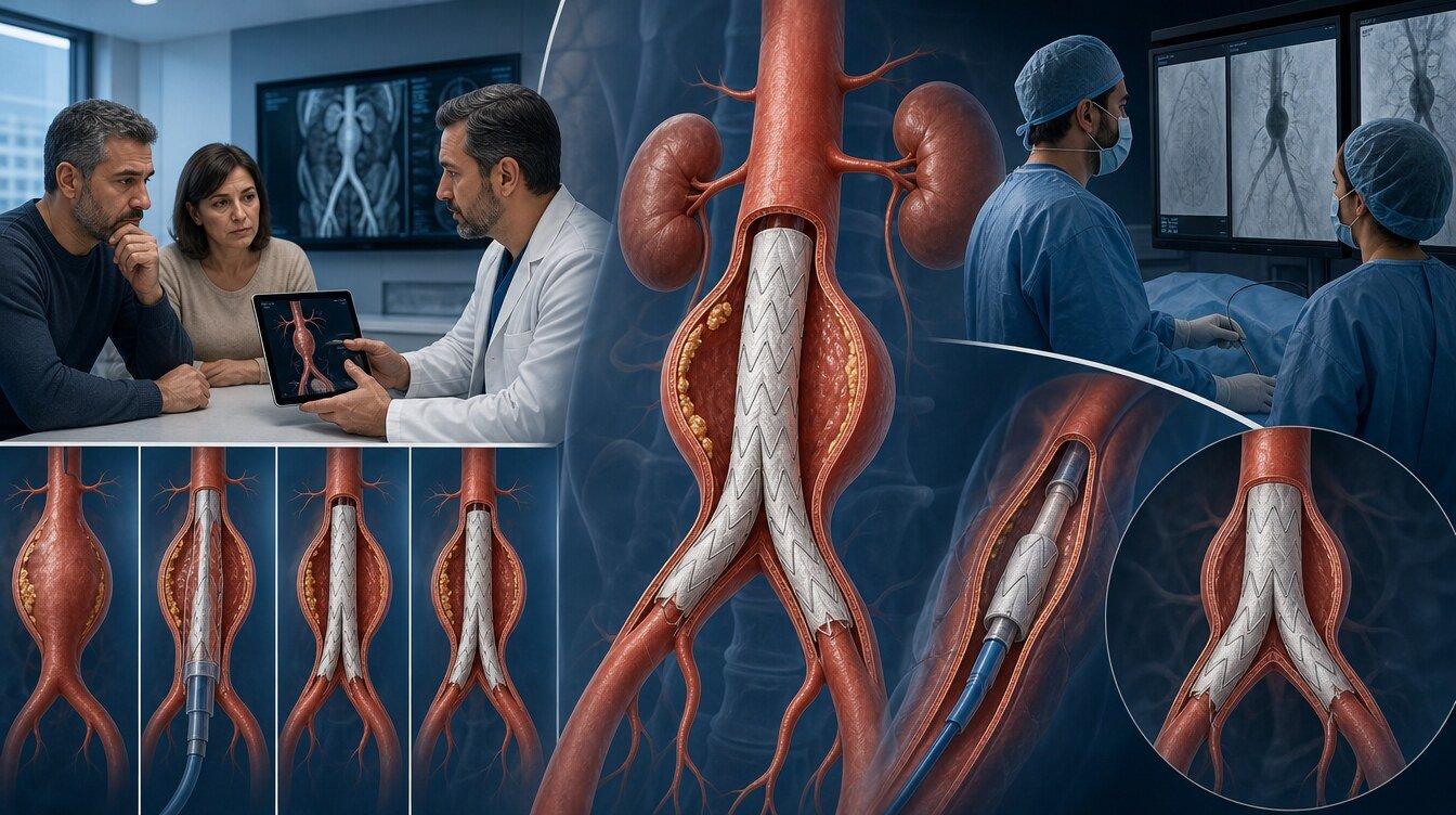
Terms used in endovascular treatment
The proximal landing zone is where the stent-graft sits on healthy aorta on the heart side of the aneurysm. The distal landing zone is the healthier seating area further down. If these zones are too short or too weak, a sealed fixation becomes harder.
Renal arteries, visceral arteries to bowel and liver, and vessels that contribute to the brain and spinal cord affect the plan. Catheters usually pass through the femoral arteries. Narrowing, calcification, tortuosity or a small calibre can make an endovascular procedure harder.
An endoleak is continued leak of blood into the aneurysm sac despite a stent-graft. Type I and III endoleaks come from the ends or junctions and can carry higher risk; they usually need treatment when found. Type II endoleak is back-filling from side branches and is the most common type. It can be watched if the sac is not growing; growth brings treatment into discussion.[5]
Reviews have reported endoleaks in about 30 percent of cases; the rate varies with the device, the definition and the length of follow-up. Some secondary procedures may be less common with modern stent-grafts than in earlier device eras, but the need for surveillance does not disappear.[5,8]
Other possible problems include migration of the stent-graft, failure of the sac to shrink or later re-growth, occlusion of a leg artery, contrast-related kidney injury, stroke, infection or bleeding, impaired spinal-cord blood flow after TEVAR and rarely paraplegia, and the need for a further endovascular or open procedure.
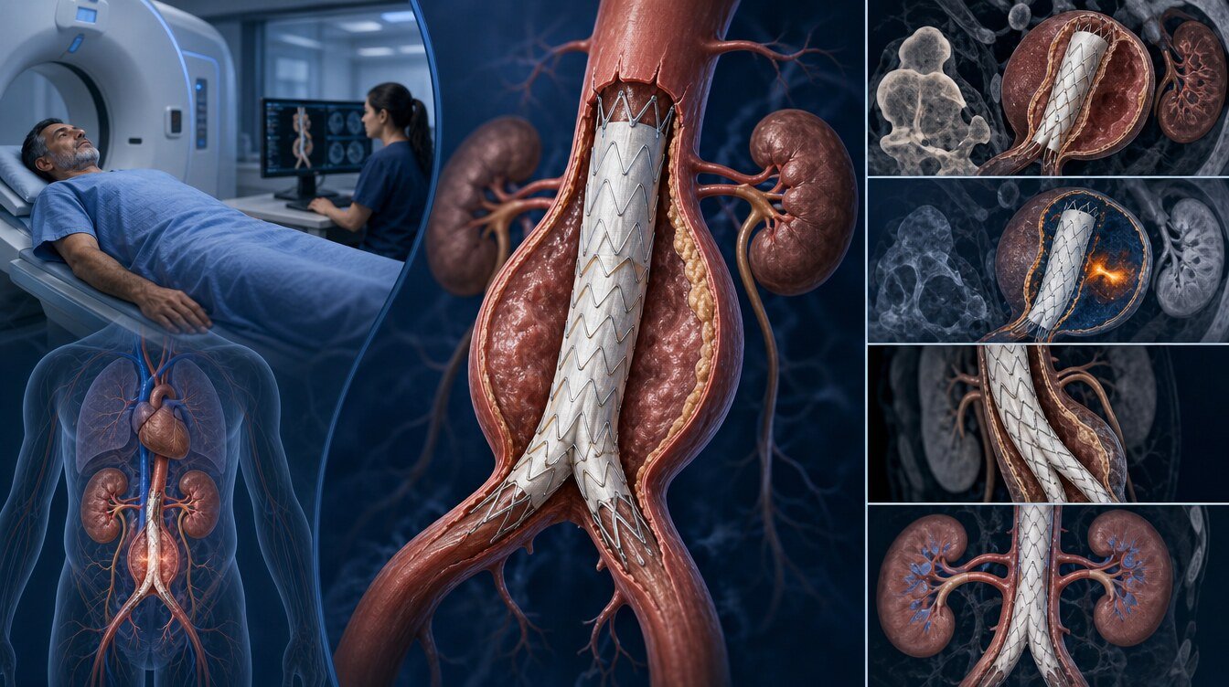
Preparation, the procedure day and recovery
The aortic team usually reviews the whole aorta and access vessels with CTA or MRA; heart, lung and kidney function; blood thinners; a plan to stop smoking and control blood pressure; infection foci; genetic counselling when suspected; and open versus endovascular options together.
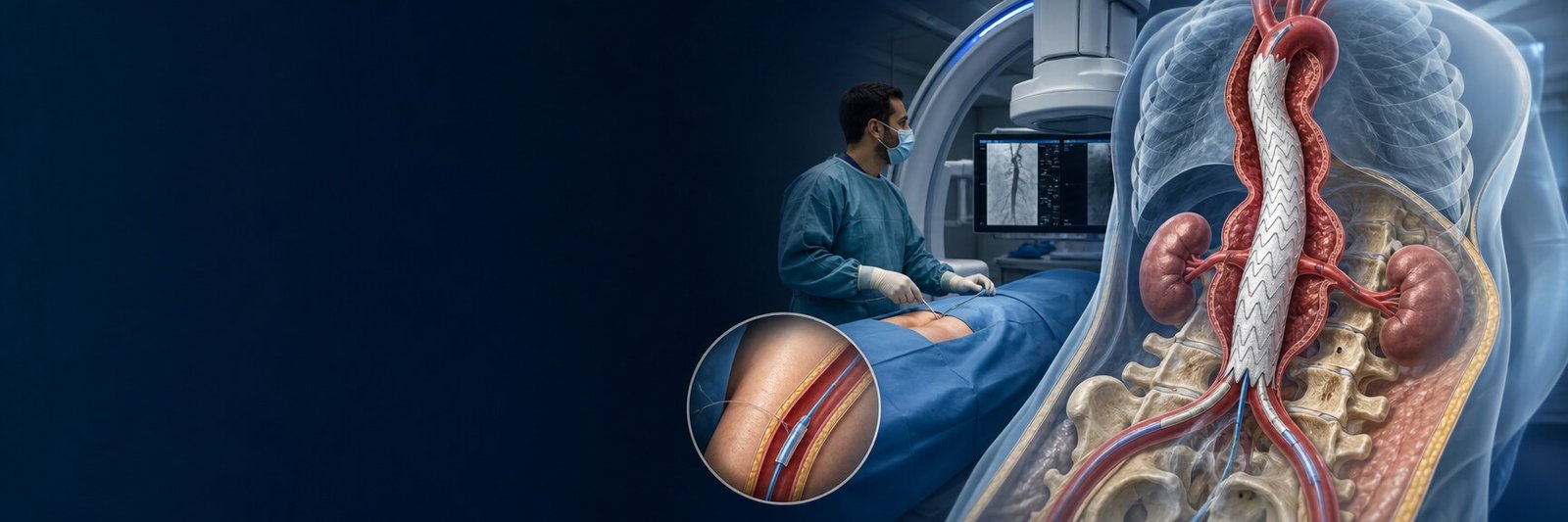
EVAR and TEVAR are usually done in an operating room or hybrid theatre under anaesthesia. Catheters enter through small groin incisions or percutaneous punctures. Under X-ray the stent-graft is advanced and opened. Technical steps differ by location; they belong on the child pages.
Length of stay depends on whether the procedure is emergency or planned, kidney and heart function, the extent of the intervention and complications. After going home, growing groin swelling or bleeding, a cold, numb or severely painful leg, fever, new chest, back, abdominal or loin pain, breathlessness, fainting or marked weakness should lead to medical care.
Who is suitable for endovascular treatment?
Suitability is not set only by being “high surgical risk” or “older”. There must be proximal and distal zones where the stent-graft can sit safely, a plan to protect branch vessels, and adequate femoral–iliac access.[1–4]
The following can make endovascular treatment harder or bring another approach into view: a very short or dilated landing zone, extremely tortuous or narrow access arteries, aortic branches inside the aneurysm, extensive thoraco-abdominal disease, genetic connective-tissue disease, an infected or inflammatory aneurysm, or a complex problem related to a previously placed stent-graft.
Open surgery, branch-preserving endovascular methods, hybrid treatment or close surveillance may then be considered. No single method is the required choice for every person.
Questions to ask an aortic team
- In which exact part of the aorta is the aneurysm?
- Is this a true aneurysm, a pseudoaneurysm or dissection-related?
- How was the diameter measured, and how much has it grown since the last imaging?
- Is surveillance safe now, and when is the next scan?
- If treatment is advised, is the aim to reduce rupture risk or to relieve symptoms?
- What are the benefits and risks of open surgery, EVAR and TEVAR for me?
- Are my landing zones, renal and other organ arteries and access vessels suitable?
- What is the risk of endoleak, stroke, kidney injury, leg occlusion or spinal-cord injury?
- Which imaging will be done after the procedure, and how often?
- What is the chance of a further procedure, and what are its warning symptoms?
- Is a genetic assessment or family screening needed?
- Would a second opinion from another aortic team be appropriate?
A segment-based decision path
The frame below does not replace a personal decision. Diameter thresholds change with the aortic segment and the type of aneurysm.
- Aortic widening is found.
- Segment and anatomy are reviewed with CTA or MRA.
- If there is rupture or shock, an emergency aortic team and emergency repair are considered.
- If there are symptoms or rapid growth, urgent specialist review and a repair plan follow.
- If diameter and risk are not near a treatment threshold, surveillance and cardiovascular-risk reduction come first.
- If repair is on the table, open surgery, EVAR or TEVAR are compared by anatomy and disease type.
- A shared, personal plan is made; regular imaging and clinical follow-up continue.
Academic context of Prof. Dr. Hakan Uçar
Among the sources that could be verified for this package, no confirmed aortic-aneurysm, EVAR or TEVAR outcome paper by Prof. Dr. Hakan Uçar was found. No specific procedure volume, device or success rate is therefore attributed to him. Coronary, TAVI or peripheral-artery papers are not used as aortic evidence.
Frequently asked questions
No. An aneurysm is weakening and widening of the aortic wall; it is not cancer. Regular assessment is still needed because of the risk of growth or rupture.
No. Many aneurysms cause no symptoms and are found on imaging done for another reason. New abdominal, back, chest or loin pain should not be ignored.
Usually not. Catheters are most often advanced from the groin arteries. Extra surgery may still be needed because of vessel anatomy or a complication.
It depends on the type. Type I and III endoleaks can carry higher risk and are usually treated. Many type II endoleaks can be watched; a procedure may be considered if the aneurysm sac grows.[5]
Iodinated contrast can worsen kidney function in some people. The risk depends on existing kidney disease, diabetes, dehydration and medicines. Kidney tests are done beforehand and precautions are taken when needed.
Complete inactivity is usually not required; the type and intensity of exercise should follow aortic diameter, blood pressure and accompanying disease. Heavy lifting or straining should be discussed with a clinician first.
A second opinion from a multidisciplinary aortic team can help when the treatment decision is borderline, genetic disease is suspected, the anatomy is complex, or the choice is between open and endovascular repair.
Literature
References
Mazzolai L, Teixidó-Turà G, Lanzi S, et al. 2024 ESC Guidelines for the management of peripheral arterial and aortic diseases. European Heart Journal. 2024;45:3538–3700. DOI: 10.1093/eurheartj/ehae179
Wanhainen A, Van Herzeele I, Bastos Goncalves F, et al. Editor's Choice — European Society for Vascular Surgery (ESVS) 2024 Clinical Practice Guidelines on the Management of Abdominal Aorto-iliac Artery Aneurysms. European Journal of Vascular and Endovascular Surgery. 2024;67:192–331. DOI: 10.1016/j.ejvs.2023.11.002
Chaikof EL, Dalman RL, Eskandari MK, et al. The Society for Vascular Surgery practice guidelines on the care of patients with an abdominal aortic aneurysm. Journal of Vascular Surgery. 2018;67:2–77.e2. DOI: 10.1016/j.jvs.2017.10.044
Upchurch GR Jr, Escobar GA, Azizzadeh A, et al. Society for Vascular Surgery clinical practice guidelines of thoracic endovascular aortic repair for descending thoracic aortic aneurysms. Journal of Vascular Surgery. 2021;73:55S–83S. DOI: 10.1016/j.jvs.2020.05.076
Smith T, Quencer KB. Best Practice Guidelines: Imaging Surveillance After Endovascular Aneurysm Repair. American Journal of Roentgenology. 2020. DOI: NA. PMID: NA.
Regalado ES, Morris SA, Braverman AC, et al. Clinical history and management recommendations of the smooth muscle dysfunction syndrome associated with ACTA2 Arg179 pathogenic variants. American Journal of Medical Genetics. 2022. DOI: NA. PMID: NA.
Rokosh RS, Wu WW, Schanzer A, Oderich GS. Society for Vascular Surgery implementation of clinical practice guidelines for patients with an abdominal aortic aneurysm. Journal of Vascular Surgery. 2021. DOI: NA. PMID: NA.
Schanzer A, Oderich GS. Management of Abdominal Aortic Aneurysms. New England Journal of Medicine. 2021;385:1690–1698. DOI: 10.1056/NEJMcp2108504
This content is for general information only and does not replace personalized medical advice. In emergencies call local emergency services.
Continue