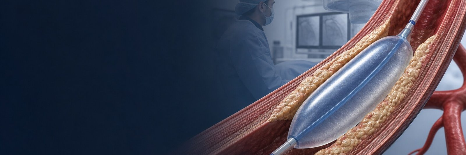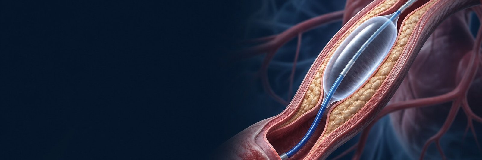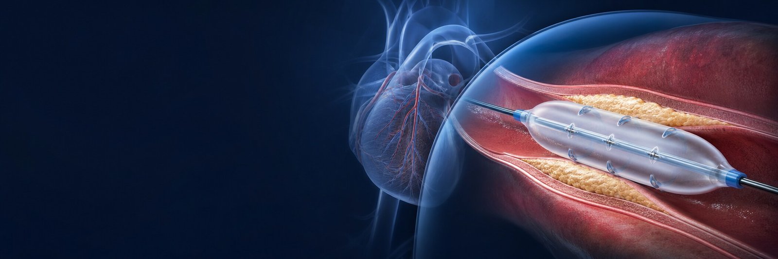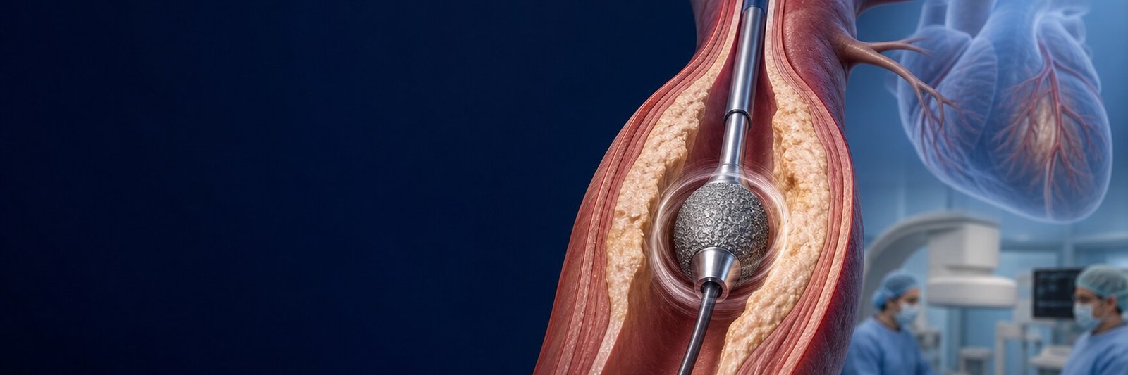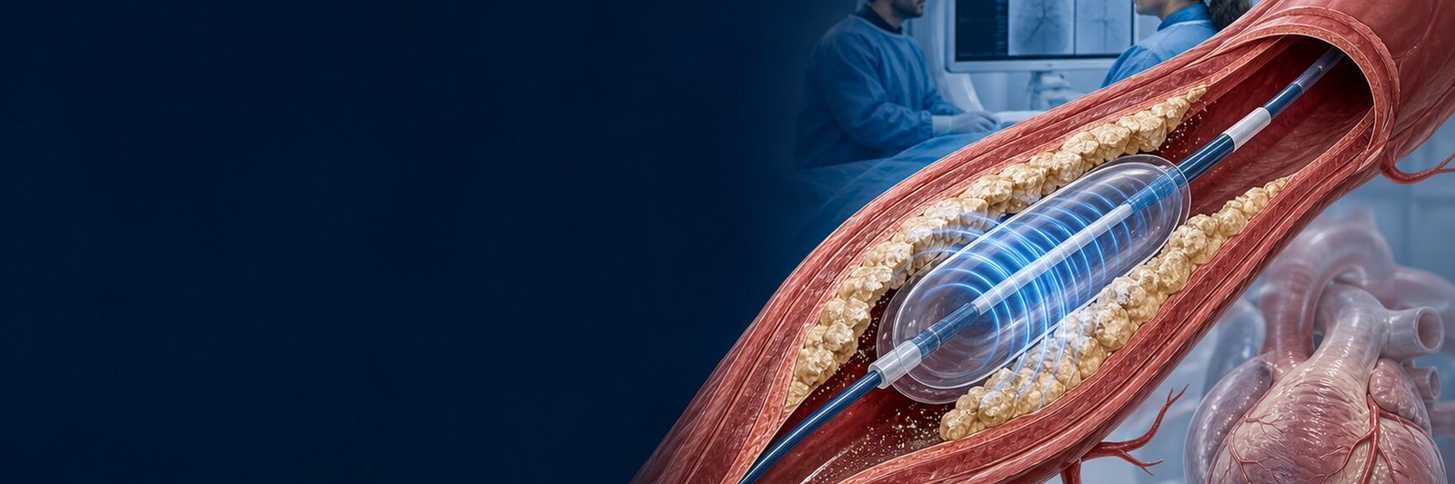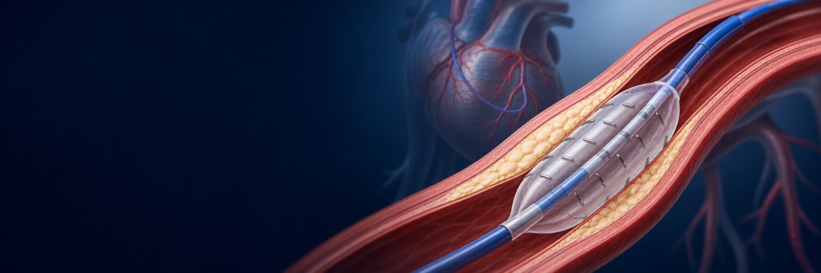
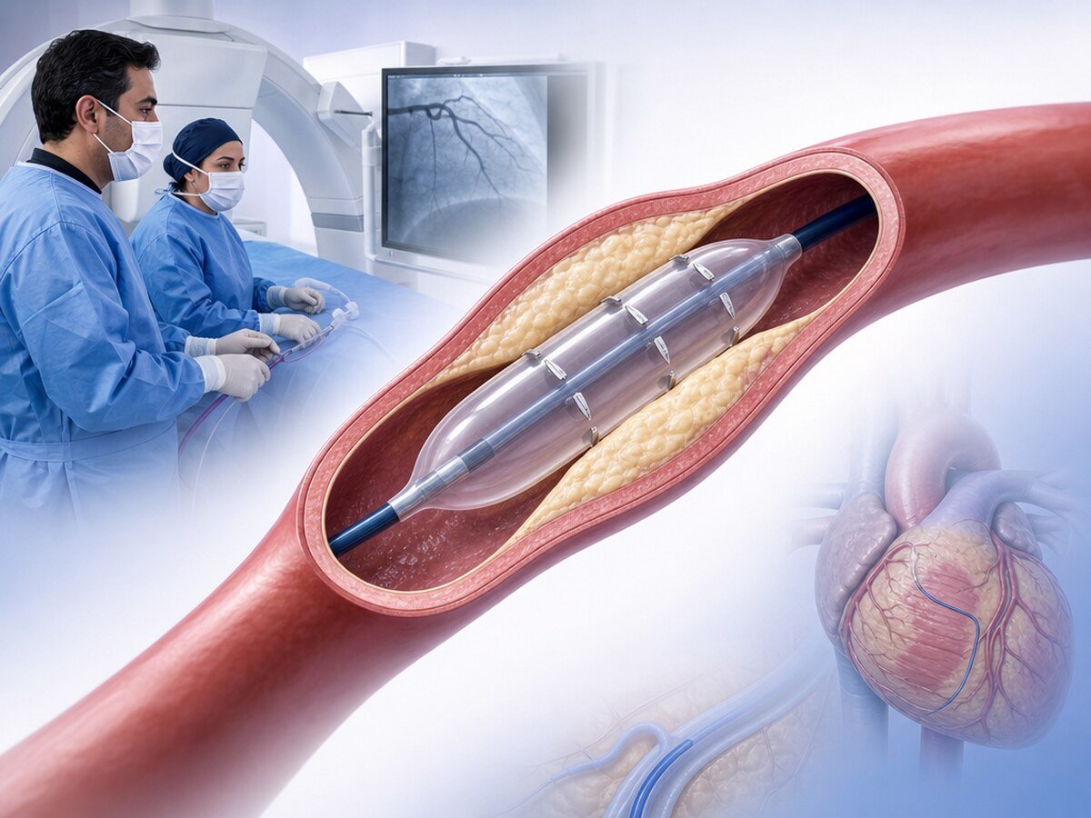
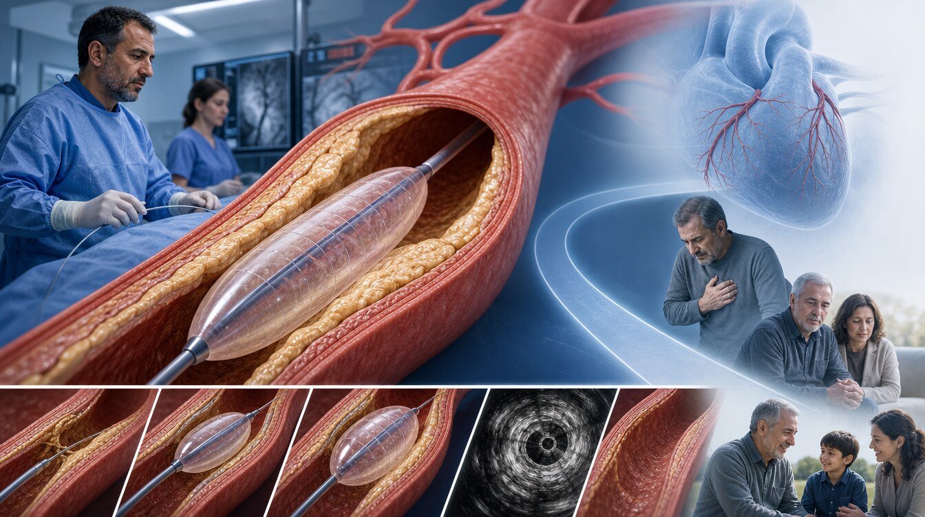
Coronary cutting balloon (microblade/atherotome balloon) is a temporary angioplasty balloon used to prepare a coronary stenosis before a stent or another treatment. Fixed longitudinal atherotomes (short microblades) on its surface aim to apply a linear effect to plaque during inflation. It is removed afterwards and is not left in the artery. It does not rotate, drill, scrape plaque or deliver a drug, and it is not required in every coronary procedure.[1]
What does a cutting balloon mean?
Cutting means linear plaque modification with fixed surface atherotomes; it does not mean an uncontrolled cut, removal of calcium or a guaranteed stent result. The aim is to help prepare the lumen in a selected stenosis. That does not mean an adequate result or fewer complications in every lesion.[1,16,17]
In everyday language, “bladed balloon” may be used once as a description of those fixed small atherotomes. It is not an uncontrolled cutting action and is not a stand-alone device-class name.
Construction, crossing profile and inflation behaviour are product-specific. COPS studied the Wolverine device; three or four surface microblades are a product-level design feature, not proof that every cutting balloon is superior.[1] This page therefore does not publish a universal pressure, diameter, duration or device sequence. A cutting balloon is not a permanent implant; it is inflated, the result is checked and it is removed with the catheter.[1]
How does a cutting balloon differ from a scoring balloon?
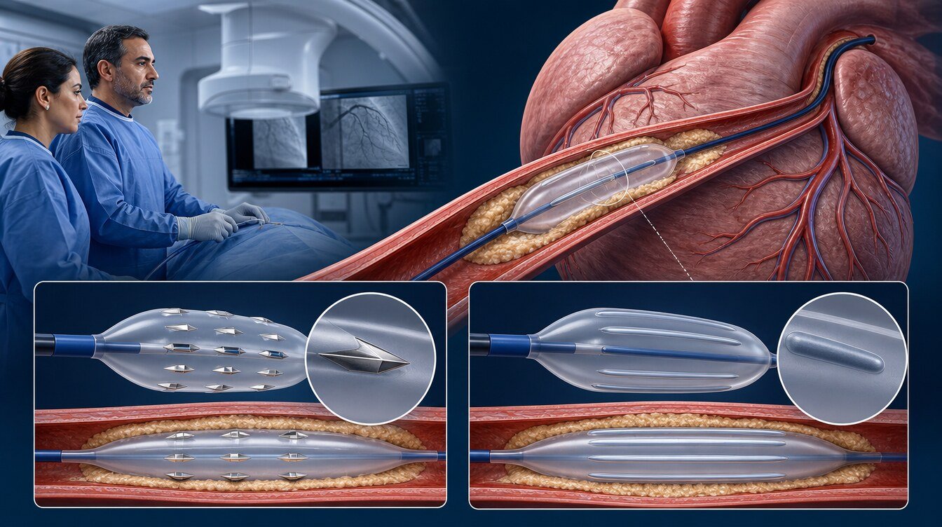
Cutting and scoring balloons may share the “modified balloon” heading; their mechanisms and clinical results must not be pooled. A cutting balloon has fixed microblades/atherotomes. A scoring balloon has surface elements (a wire, a helical nitinol element or nylon ridges) that aim to concentrate force; those elements are not blades.[16,17]
Pooled analyses may place cutting and scoring in the same bucket. That does not make the tools the same or allow one result to be transferred to the other.[13]
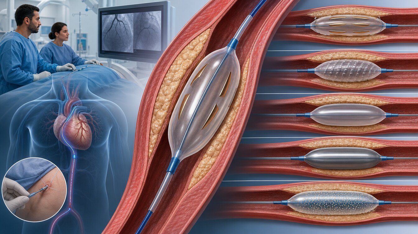
How does it differ from other balloons and methods?
More than one tool may be used in the same procedure. That does not make the categories interchangeable or give them the same evidence.
| Feature | Cutting balloon | Scoring balloon | Standard NC balloon | Super high-pressure NC (e.g. OPN NC) | Drug-coated balloon (DCB) |
|---|---|---|---|---|---|
| Main purpose | Linear plaque modification with microblades | Focused preparation via surface elements | Controlled mechanical expansion | Selected resistant lesions not opened by a standard NC balloon | Drug transfer to the vessel wall |
| Remains in the artery? | No | No | No | No | No |
| Removes calcium? | No | No | No | No | No |
| Delivers drug? | No | No | No | No | Yes |
| Needed in every patient? | No; by lesion and result | No | No | No; a separate device class | No |
Intravascular lithotripsy (IVL) uses acoustic shockwaves and rotational atherectomy uses debulking/ablation. A cutting balloon is not a universal substitute. Peripheral balloons are outside this coronary evidence.[16,17]
When may it be considered?
A cutting balloon is not a routine step before every stent or in every calcified stenosis. Use depends on whether the lesion can be crossed, calcium morphology, the stent or drug-coated balloon plan and, when used, intravascular imaging.[16,17]
Calcified de novo stenoses
In a new (de novo) calcified stenosis a cutting balloon may be one preparation option before stenting. COPS examined an imaging endpoint in selected patients with IVUS-confirmed calcification; it was not powered to show fewer deaths, myocardial infarctions or repeat procedures. Uncrossable lesions or those not opened by a standard NC balloon may need other tools.[1,16,17]
In-stent restenosis (historical context)
In the older PTCA/BMS era, cutting balloons were studied for in-stent restenosis (ISR). RESCUT and the small Montorsi trial belong to that period. Procedural or imaging observations such as less slippage or less early lumen loss must not be transferred to modern drug-eluting-stent ISR (DES-ISR) or drug-coated balloon care.[2,8]
Preparation before a drug-coated balloon
A cutting balloon may be used before a drug-coated balloon (DCB); it is not itself a drug-coated balloon. No completed human cutting-specific comparative DCB superiority was verified in this package. NATURE and SCORE-CUT are design records intended to address that question; they are not outcome reports.[19,20]
When may it be insufficient alone?
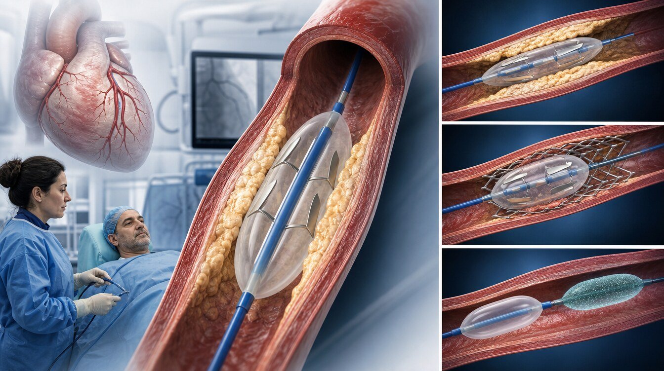
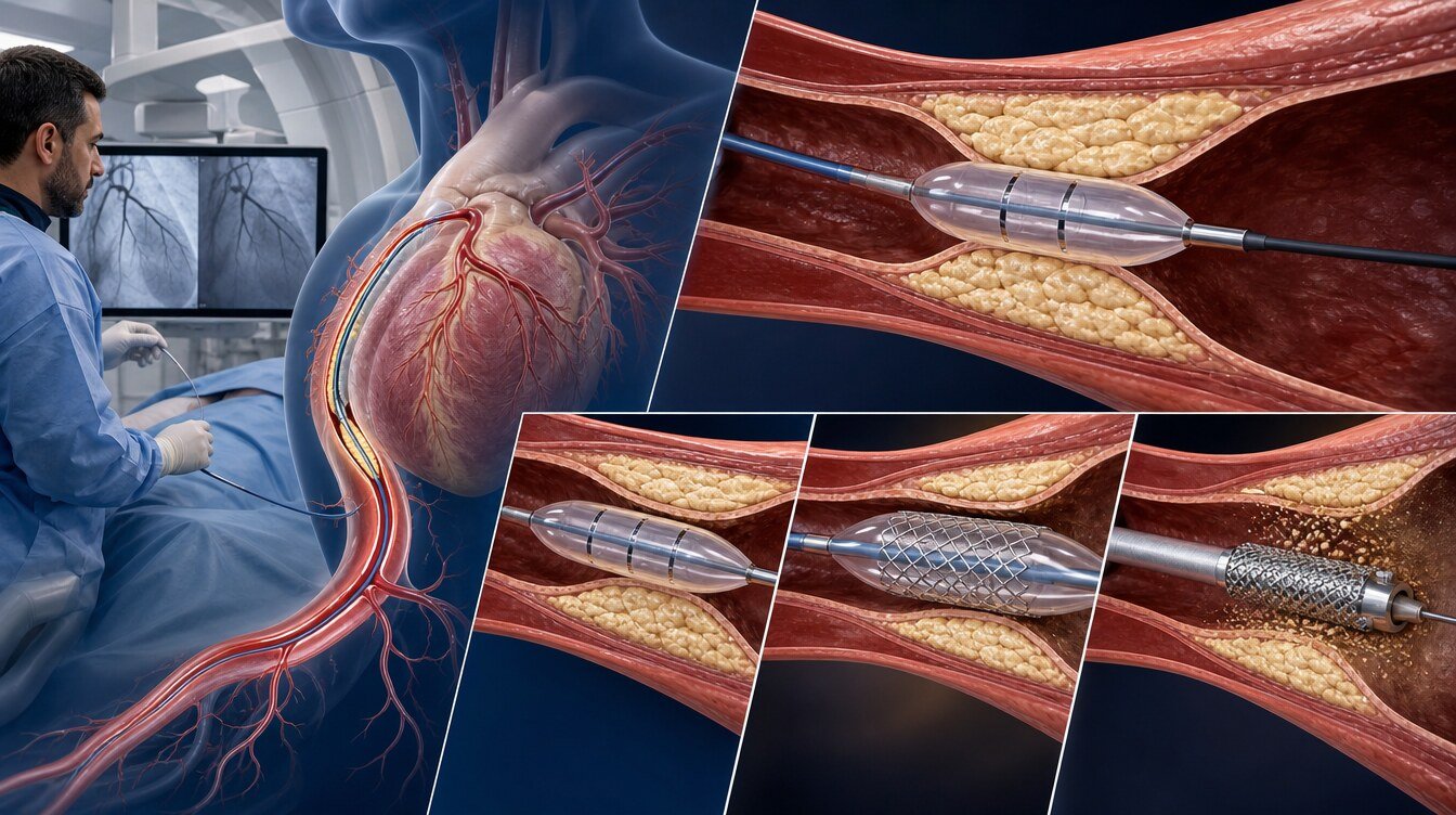
A cutting balloon may not be enough in deep or circumferential calcium, uncrossable lesions, under-expanded stents or when another mechanism dominates. Consensus texts note that other methods may be needed according to calcium phenotype; a single balloon is not a guaranteed solution.[16,17]
A different strategy may be needed when:
- The balloon cannot cross the stenosis
- The balloon crosses but cannot expand the lesion adequately
- Standard NC or cutting preparation remains inadequate
- Deep, long or circumferential calcium needs another modification method
- Calcium beneath a stent resists balloon force
- Dissection or flow-limiting vessel injury occurs during the procedure
A scoring balloon, a standard or super high-pressure NC balloon, rotational atherectomy, IVL or a combined approach may then be considered. These tools are not universal substitutes for one another. Adding a cutting balloon after rotational atherectomy also does not always mean better stent expansion.[12,16,17]
Why may IVUS or OCT be used?
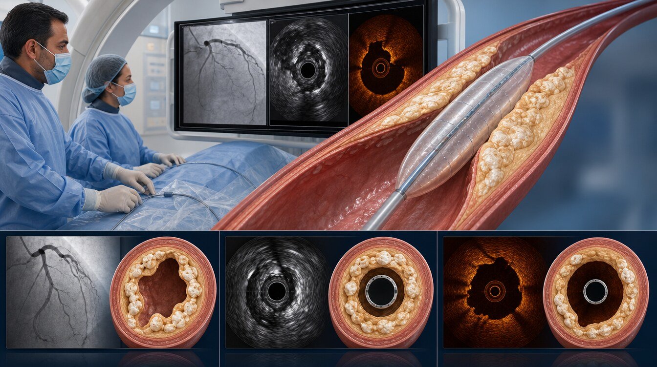
Coronary angiography gives a two-dimensional lumen view and can miss calcium depth, circumferential extent and true stent expansion. Intravascular ultrasound (IVUS) and optical coherence tomography (OCT) provide cross-sectional images that can help assess whether calcium is superficial or deep, whether a balloon or stent has expanded, whether the stent is apposed, and the mechanism of restenosis.[16,17]
SCAI and EAPCI consensus documents separate cutting and scoring designs and discuss intravascular imaging as a guide to calcium phenotype. That does not mean imaging prevents heart attack or death, that it mandates a cutting balloon, or that it is technically possible in every patient. No universal expansion cut-off is published here.[16,17]
How is the procedure performed?
The procedure is done in a catheter laboratory equipped for coronary angiography and PCI. It is not a separate operation unique to the cutting balloon; the patient experience is similar to other coronary balloon-stent procedures:[21]
- Preparation: History, medicines, blood tests, kidney function and contrast risk are reviewed.
- Access: Radial or femoral artery under local anaesthesia.
- Imaging and wire: The artery is imaged; a fine wire is advanced beyond the stenosis.
- Preparation and treatment plan: The team chooses a cutting balloon, NC balloon, scoring balloon, stent, drug-coated balloon or another tool according to the lesion and the live result.
- Result check: Flow, residual stenosis and, when used, IVUS/OCT findings are reassessed.
- Removal: The cutting balloon is removed. If a stent was placed, the lasting structure is the stent.
Most patients remain awake. Brief chest discomfort during inflation should be reported if new or severe.[21]
This page does not prescribe pressure, inflation time, balloon diameter or device order.
What does the scientific evidence show?
Three ideas should be kept apart. Device success is a protocol-defined step such as delivery, inflation and adequate preparation without rupture or perforation. Strategy success is whether the chosen plan—including a bailout tool if needed—treats the lesion adequately. Bailout is a switch to another device when the planned tool is not enough. These are not the same.[1,9]
Contemporary calcified lesions: COPS
COPS was a multicentre open-label trial comparing a Wolverine cutting balloon with a standard NC balloon in severely calcified de novo lesions with an IVUS calcium arc of at least 100 degrees (100 randomised; 87 per-protocol). The primary endpoint was minimum stent area at the calcified segment (an imaging endpoint). In the per-protocol analysis the cutting arm had a larger final lumen area and larger, more symmetric stent expansion. One-year clinical event counts were sparse; the trial was not powered to show fewer deaths, myocardial infarctions or repeat procedures.[1]
COPS defined device success as delivery, inflation, adequate lesion preparation and no vessel rupture or perforation. The report also states that all lesions were successfully treated with the study device and that no additional device was required, yet it records three cutting-balloon device failures: one lesion needed rotational atherectomy to advance the cutting balloon (the cutting balloon was still used) and two Ellis type-1 perforations. “All lesions were uncomplicated device successes” is therefore not a fair summary. Final strategy success was not separately defined and is internally ambiguous in the accessible text.[1]
Cutting after rotational atherectomy: ROTA-CUT
ROTA-CUT compared rotational atherectomy followed by a cutting balloon with rotational atherectomy followed by an NC balloon in 60 patients. IVUS minimum stent area was similar (about 6.7 versus 6.9 mm²). Adding a cutting balloon after rotational atherectomy did not increase stent area versus an NC balloon. Follow-up is short; a universal “RA plus cutting is required” ranking cannot be set.[12]
The PREPARE-CALC family (mixed modified balloon)
PREPARE-CALC compared rotational atherectomy with a mixed modified-balloon strategy (scoring and cutting combined) in 200 severely calcified native lesions. Strategy success was 81% with modified balloons versus 98% with rotational atherectomy. Nine-month late lumen loss and reported clinical outcomes were similar. The two- and five-year papers are follow-up of the same 200-patient cohort; at five years target-vessel failure was comparable and target-lesion revascularisation was lower after rotational atherectomy. This family does not yield a cutting-specific randomised estimate.[9–11]
Older PTCA/BMS-era evidence (historical boundary)
The Cutting Balloon Global Randomized Trial compared a cutting balloon with contemporary PTCA in 1,238 de novo lesions. Six-month angiographic restenosis was not reduced; five perforations occurred in the cutting arm. This is not DES/DCB-era evidence.[3]
CAPAS was a historical PTCA trial using lesion-level randomisation in small-vessel type B/C lesions; it cannot be transferred to current practice. A PMID was not verified in this package.[4]
REDUCE III compared cutting-balloon preparation before a bare-metal stent with balloon-BMS in 521 patients and reported lower BMS-era restenosis/TLR. That is not a modern drug-eluting-stent comparison. A PMID was not verified in this package.[6]
RESCUT compared a cutting balloon with conventional PTCA in 428 patients with ISR. Balloon slippage was lower with cutting; recurrent binary restenosis at seven months (29.8% versus 31.4%) and clinical events were similar. This is historical PTCA/BMS ISR care and must not be transferred to current DES-ISR/DCB treatment.[2]
Montorsi and colleagues randomised 50 ISR patients to cutting balloon or PTCA; immediate lumen change was similar, while 24-hour early loss and observed TLR were lower with cutting. It is a small, single-centre historical ISR/IVUS study. A DOI was not verified in this package.[8]
Umeda and Tang appear in the 2024 meta-analysis reference list. Detailed primary fields were not verified in this package, so they are not used for numerical clinical claims.[5,7]
Pooled analyses and ongoing records
The 2024 native-vessel meta-analysis pools cutting and scoring balloons and mixes POBA, BMS and DES strategies. Pooled target-lesion revascularisation was lower; the authors highlight a small, often non-inferiority-oriented evidence base with a historical POBA component. The article prose cites eight studies and 2,712 patients, while the forest plot names seven analysable lines. This cannot establish a cutting-specific contemporary patient-important benefit.[13]
NATURE is a 200-patient randomised design record comparing a cutting balloon with a standard balloon before DCB in de novo lesions. SCORE-CUT is a design record comparing NC, scoring and cutting strategies before DCB. Neither is outcome evidence.[19,20]
What are possible benefits and limitations?
| Possible benefit / purpose | Limitation / required caveat |
|---|---|
| May help preparation of a selected stenosis via linear atherotome effect | Does not clear calcium or guarantee stent expansion |
| In COPS, the imaging endpoint favoured cutting in IVUS-selected calcified lesions | Small trial; not evidence of fewer deaths/MI/repeat procedures; device-success wording is internally inconsistent |
| RESCUT showed less balloon slippage | Historical ISR era; recurrent restenosis and clinical events were similar |
| Balloon is removed and adds no new permanent implant | Most de novo plans still include a stent |
| May be used before a DCB | Does not deliver drug; cutting-specific DCB superiority has not been shown |
What are the risks?
A cutting balloon is used in coronary intervention. Risks of balloon inflation overlap with PCI generally:[1,3,21]
- Coronary dissection
- Coronary perforation
- Failure to cross, device entrapment or balloon damage
- Slow flow or no-reflow
- Acute vessel closure
- Side-branch compromise
- Myocardial infarction, stent thrombosis or arrhythmia
- Access-site bleeding and vascular complications
- Contrast-related kidney injury or allergy
- Later restenosis if a stent is used
The two Ellis type-1 perforations in the COPS cutting arm and the five perforations in the Global Trial must not be copied as a personal risk calculator. Small-trial events are not universal rates.[1,3]
What are the alternatives?
| Method | Core approach | Key difference from a cutting balloon |
|---|---|---|
| Scoring balloon | Focused preparation via surface elements | No microblades; evidence is not transferable |
| Standard NC balloon | Controlled mechanical expansion without surface elements | Cutting is not “a better NC balloon” |
| Super high-pressure NC (e.g. OPN NC) | Twin-layer resistant-lesion device | A separate device class |
| Drug-coated balloon (DCB) | Antiproliferative drug transfer | Cutting does not deliver drug; it may prepare a stenosis before DCB |
| Rotational atherectomy | Debulks hard tissue to create a path | Different mechanism; ROTA-CUT found no MSA gain versus NC after RA |
| Intravascular lithotripsy (IVL) | Pressure waves that fracture calcium | Different mechanism; the balloon must still cross |
Methods may be combined; combined use is not routine for every patient.[16,17]
Preparation, discharge and recovery
Preparation depends on elective versus urgent setting, kidney function, bleeding risk, medicines, contrast history and access route. Follow hospital fasting and medicine instructions; do not stop prescribed medicines on your own.[21]
After planned uncomplicated coronary angioplasty some patients go home the same or next day. Local policy, clinical status and complications change the timing. This general frame comes from NHS patient information and is not a fixed discharge rule for every hospital in Türkiye.[21]
Discharge advice should be personalised and include access-site care, activity limits, antiplatelet and other prescriptions, follow-up, and when to seek emergency care.
Cutting-balloon use alone does not set antiplatelet duration. The plan depends on stenting, a drug-coated balloon, the clinical syndrome and bleeding–clotting risk. Stopping antiplatelets without advice can cause serious stent thrombosis.[14,21]
Bruising at the access site can occur. Increasing pain, swelling, redness, discharge or fever should prompt contact with the treating centre or another healthcare service.[21]
When should emergency services be called?
Call emergency services (112 in Türkiye) without delay for:[21]
- Chest pain that does not settle or is worsening
- Access-site bleeding that continues despite direct pressure or starts again
- Marked coldness, colour change or numbness in the treated arm or leg
- Fainting, severe breathlessness, sudden weakness or a change in consciousness
Not every bruise or mild tenderness is an emergency; uncontrolled bleeding, severe chest pain or impaired limb circulation is. Do not drive yourself with emergency symptoms. The 112 direction is a general safety frame; detailed local discharge wording should be reviewed against an official Turkish source before publication.[21]
Frequently asked questions
No. The preferred name is coronary cutting balloon (microblade/atherotome balloon). “Bladed balloon” is only a lay description of the fixed surface atherotomes; it does not mean an uncontrolled cut or a separate device class.[1]
No. COPS is a small trial with an imaging primary endpoint. Device success, strategy success and bailout are separate ideas. A larger stent area is not a reduction in death or myocardial infarction.[1]
No. RESCUT is a historical PTCA/BMS-era ISR trial; recurrent restenosis and clinical events at seven months were similar. It must not be transferred to modern DES-ISR/DCB treatment.[2]
No. In ROTA-CUT, adding a cutting balloon after rotational atherectomy did not increase IVUS minimum stent area versus an NC balloon. A small trial does not set a universal ranking.[12]
No. The PREPARE-CALC modified-balloon arm combined scoring and cutting balloons; it is not cutting-specific evidence. Strategy success was lower than with rotational atherectomy.[9]
Assessment and decision
A cutting balloon is a temporary preparation tool used in selected coronary procedures. Planning is broader than “will a cutting balloon be used?” and includes treatment need, device crossability, calcium morphology, the stent or drug-coated balloon plan, IVUS/OCT findings and alternatives.[14–18]
The 2021 ACC/AHA/SCAI guideline, the 2024 ESC guideline, the 2023 EAPCI consensus, the 2024 SCAI calcified-lesion consensus and the SCAI ISR statement have no cutting-specific Class/LOE recommendation verified in this package; the decision rests on individual clinical assessment.[14–18]
Literature
References
Mangieri A, et al. Cutting balloon to optimize predilation for stent implantation: The COPS randomized trial. Catheter Cardiovasc Interv. 2023. DOI: 10.1002/ccd.30603. PMID: 36841945.
Albiero R, et al. Cutting balloon versus conventional balloon angioplasty for the treatment of in-stent restenosis: results of the RESCUT trial. J Am Coll Cardiol. 2004. DOI: 10.1016/j.jacc.2003.09.054. PMID: 15028348.
Mauri L, et al. Cutting balloon angioplasty for the prevention of restenosis: results of the Cutting Balloon Global Randomized Trial. Am J Cardiol. 2002. DOI: 10.1016/S0002-9149(02)02773-X02773-X). PMID: 12423707.
Izumi M, et al. Final results of the CAPAS trial. Am Heart J. 2001. DOI: 10.1067/mhj.2001.119129. A PMID was not verified in this package.
Umeda H, et al. Promising efficacy of primary gradual and prolonged balloon angioplasty in small coronary arteries. Am Heart J. 2004. DOI: 10.1016/j.ahj.2003.07.016. Detailed primary fields and a PMID were not verified in this package; not used for numerical clinical claims.
Ozaki Y, et al. Impact of cutting balloon angioplasty prior to bare metal stenting on restenosis. Circ J. 2007. DOI: 10.1253/circj.71.1. A PMID was not verified in this package.
Tang Z, et al. Cutting-balloon angioplasty before drug-eluting stent implantation for the treatment of severely calcified coronary lesions. J Geriatr Cardiol. 2014. DOI: 10.3969/j.issn.1671-5411.2014.01.012. Primary-report details were not verified in this package; not used for numerical clinical claims.
Montorsi P, et al. Randomized trial of conventional balloon angioplasty versus cutting balloon for in-stent restenosis. 2004. PMID: 15185885. A DOI was not verified in this package.
Abdel-Wahab M, et al. High-Speed Rotational Atherectomy Versus Modified Balloons Prior to Drug-Eluting Stent Implantation in Severely Calcified Coronary Lesions: The Randomized PREPARE-CALC Trial. Circ Cardiovasc Interv. 2018. DOI: 10.1161/CIRCINTERVENTIONS.118.007415. Trial: NCT02502851. A PMID was not verified in this package.
Allali A, et al. High-speed rotational atherectomy versus modified balloons for plaque preparation of severely calcified coronary lesions: two-year outcomes of the randomised PREPARE-CALC trial. EuroIntervention. 2022. DOI: 10.4244/EIJ-D-22-00677. PMID: 36579635. PMCID: PMC10068860. Trial: NCT02502851.
Mankerious N, et al. Lower revascularization rates after high-speed rotational atherectomy compared to modified balloons in calcified coronary lesions: 5-year outcomes of the randomized PREPARE-CALC trial. Clin Res Cardiol. 2024. DOI: 10.1007/s00392-024-02434-1. PMID: 38483633. Trial: NCT02502851.
Sharma SK, et al. Rotational atherectomy combined with cutting balloon to optimise stent expansion in calcified lesions: the ROTA-CUT randomised trial. EuroIntervention. 2024. DOI: 10.4244/EIJ-D-23-00811. PMID: 38165112. PMCID: PMC10756220.
Sorolla Romero JA, et al. Use of cutting or scoring balloons in patients with native coronary artery disease: systematic review and meta-analysis. REC Interv Cardiol. 2024. English DOI: 10.24875/RECICE.M24000448. Spanish DOI: 10.24875/RECIC.M24000450. PMID: 40415775. PMCID: PMC12097377. Registration: CRD42023434007.
Lawton JS, et al. 2021 ACC/AHA/SCAI Guideline for Coronary Artery Revascularization. Circulation. 2022. DOI: 10.1161/CIR.0000000000001038. No cutting-specific Class/LOE was verified in this package.
Vrints C, et al. 2024 ESC Guidelines for the management of chronic coronary syndromes. Eur Heart J. 2024. DOI: 10.1093/eurheartj/ehae177. No cutting-specific Class/LOE was verified in this package.
Barbato E, et al. Management strategies for heavily calcified coronary stenoses: an EAPCI clinical consensus statement in collaboration with the EURO4C-PCR group. Eur Heart J. 2023. DOI: 10.1093/eurheartj/ehad342.
Riley RF, et al. SCAI Expert Consensus Statement on the Management of Calcified Coronary Lesions. JSCAI. 2024. DOI: 10.1016/j.jscai.2023.101259. PMID: 39132214. PMCID: PMC11307856.
Klein LW, et al. SCAI Expert Consensus Statement on Management of In-Stent Restenosis and Stent Thrombosis. JSCAI. 2024. DOI: 10.1016/j.jscai.2023.101626. No cutting-specific Class/LOE was verified in this package.
Ono M, et al. Cutting balloon versus standard balloon for lesion preparation of drug-coated balloon treatment in de-novo coronary artery lesions: Rationale and Design of the Randomized NATURE trial. Cardiovasc Revasc Med. 2025. Trial: jRCTs032230543. DOI/PMID were not verified in this package; this is not an outcome report.
Khandkar C, et al. SCORE-CUT: A Randomized Trial Comparison of Scoring, Cutting, and Non-Compliant Balloon Lesion Preparation Strategies for Drug-Coated Balloon Angioplasty—Rationale and Design. Catheter Cardiovasc Interv. 2026. Trial: ACTRN12625001349437. DOI/PMID were not verified in this package; this is not an outcome report.
NHS. Coronary angioplasty — Recovery. Official patient information. Accessed 27 August 2026.
Continue