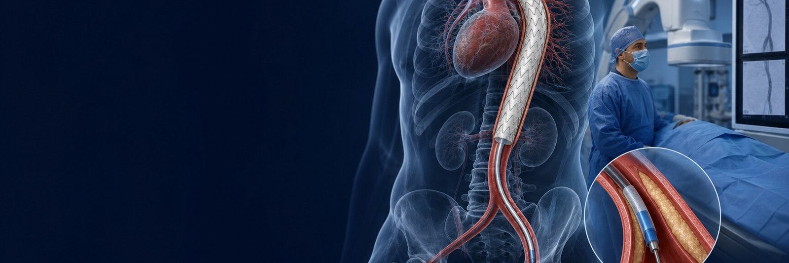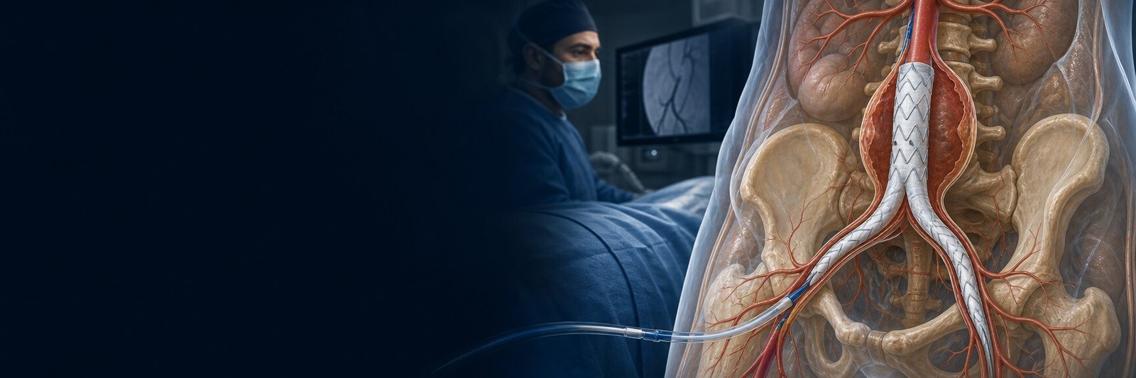
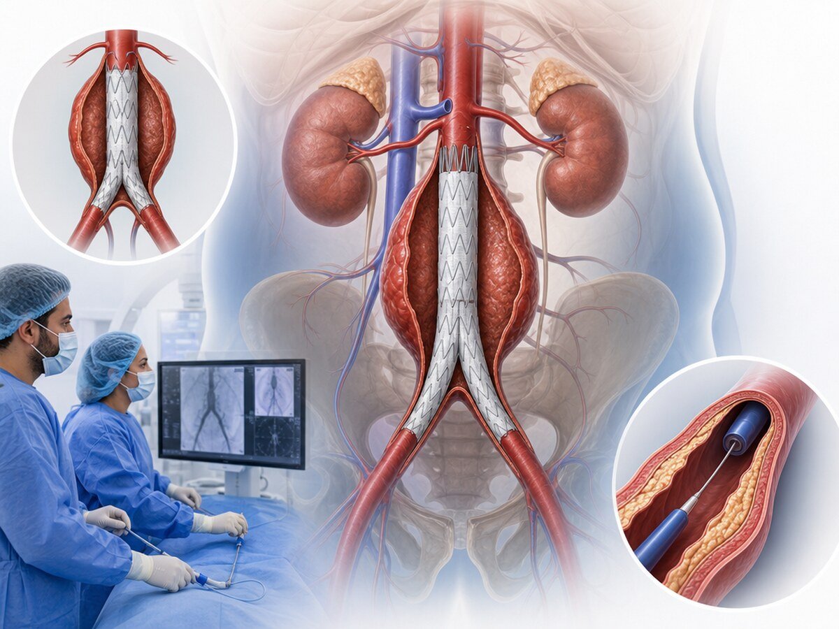
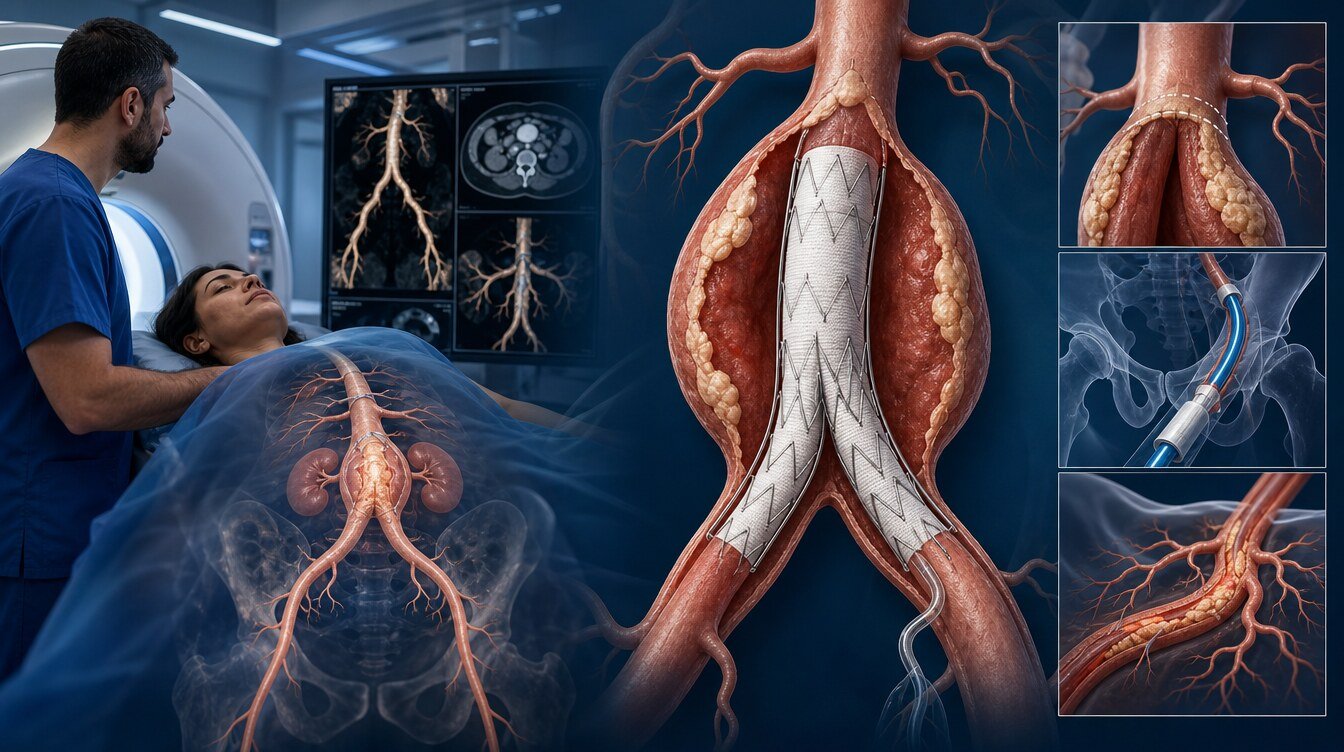
EVAR is short for endovascular aneurysm repair. A covered vessel prosthesis — a stent-graft — is advanced from the groin arteries into an abdominal aortic aneurysm. The prosthesis aims to take blood flow away from the inside of the aneurysm sac and reduce pressure on the aortic wall.[1,2]
EVAR can be used for the part of the abdominal aorta below the renal arteries and for some aorto-iliac aneurysms. Not every person or every vessel anatomy is suitable; the decision should be made by an Aortic Team after CT images, general health and a comparison with open surgery are reviewed.[1,2]
This article is a child page of aortic aneurysm and endovascular treatment. TEVAR, endovascular treatment of the thoracic aorta, is outside the scope of this page; TEVAR indications and outcome rates are not transferred to EVAR.
What is an abdominal aortic aneurysm?
The aorta is the largest artery leaving the left ventricle. The part inside the chest is the thoracic aorta; the part inside the abdomen is the abdominal aorta.
An aneurysm is widening of a vessel beyond its usual size because the wall has weakened. Lasting widening of the abdominal aorta is an abdominal aortic aneurysm (AAA). As it grows, wall tension can rise. A markedly enlarged aneurysm can tear and cause serious bleeding into the abdomen or surrounding tissues. That is called rupture.[1,2]
An infrarenal aneurysm sits below the renal arteries, in the lower abdominal aorta. Standard EVAR is most often considered for this region. An aorto-iliac aneurysm extends from the abdominal aorta into the iliac arteries. Patency of the iliac arteries and whether the internal iliac arteries that supply pelvic organs can be preserved are then planned separately. More complex aneurysms that involve the renal or mesenteric arteries may need fenestrated or branched stent-grafts.[2]
Clinical states of the aneurysm
A solid, asymptomatic (intact) aneurysm that has not ruptured and is not causing symptoms may be repaired in a planned, elective way.
A symptomatic aneurysm may be suggested by new or unexplained abdominal, loin or flank pain, tenderness related to the aneurysm or pressure on surrounding tissues. Urgent assessment can be needed even without rupture.
A leaking aneurysm means a small tear has formed and blood is leaking in a limited way into the surroundings; this can be a contained rupture. The person may still look stable at first, but the picture can worsen quickly.
A ruptured aneurysm with active internal bleeding is a ruptured abdominal aortic aneurysm and is life-threatening. If the anatomy is suitable, an emergency endovascular strategy can be considered; if it is not, open repair may be needed.[6]
Screening, diagnosis and EVAR planning
Screening looks for disease before symptoms appear. Ultrasound is usually used to screen for abdominal aortic aneurysm.
The US Preventive Services Task Force (USPSTF) recommends a one-time ultrasound screen in men aged 65–75 who have ever smoked. In men of the same age who have never smoked, screening can be considered according to personal risk. Routine screening is not recommended in women who have never smoked and have no family history of AAA; in women with a smoking or family history the evidence does not allow a firm conclusion. Recommendations vary by country and health system.[7]
Ultrasound can show that an aneurysm is present and its approximate diameter. Most people need CT angiography (CTA) when EVAR is planned. CTA is used to review maximum diameter and length; the proximal neck below the renal arteries; neck length, diameter, thrombus or calcium; angulation; iliac diameter, tortuosity, narrowing and calcium; the route from the groin; branches to the kidneys, bowel and pelvic organs; and where the distal ends of the stent-graft can sit.[2,3]
Diameter and growth rate matter for the treatment decision. Current guidelines may bring repair into discussion around about 5.0–5.5 cm in men and about 5.0 cm in women; these values are not an automatic operation limit. Symptoms, rapid growth, shape, rupture risk, life expectancy and procedural risk can change the decision.[1,2]
Why is not everyone suitable for EVAR?
Each stent-graft has an anatomical range of use set out in its instructions for use (IFU). A very short, highly angulated, thrombus-lined or irregular neck; very narrow or tortuous iliac arteries; a risk of covering important branches; and aorto-iliac extension can make standard EVAR less suitable.[2,3]
The question is therefore not only “is the diameter enough?” but also “can the stent-graft sit safely and stay in place over the years?”
Treatment options
The choice brings together diameter and growth, symptoms, rupture status, anatomy, age, heart, lung and kidney disease, life expectancy and the person’s preferences. No method is universally superior.[1,2]
| Option | Basic approach | Usually considered when | Important points |
|---|---|---|---|
| Surveillance and risk reduction | Imaging at intervals; stopping smoking and treating cardiovascular risk | Small, asymptomatic aneurysms that do not need immediate repair | The plan can change if the aneurysm grows or symptoms appear |
| Standard EVAR | Stent-graft placement from the groin arteries | Anatomy fits the device instructions for use | Can be less invasive; lifelong imaging and sometimes a further procedure are needed |
| Fenestrated or branched EVAR | Stent-grafts with special openings or side branches for renal or mesenteric arteries | Complex abdominal or thoraco-abdominal aneurysms | Planning and follow-up can be more complex |
| Open repair | Surgical replacement of the aneurysmal segment with a prosthetic graft | EVAR anatomy is not suitable, or in some younger people with a long life expectancy | A larger surgical load; durability and early risk are weighed together |
During surveillance, blood pressure, cholesterol, diabetes and other arterial diseases are managed. These steps do not remove the existing aneurysm.[1]
How is EVAR done?
The Aortic Team reviews the CTA and plans the size and position of the stent-graft. Heart, lung and kidney status, current medicines, blood thinners and previous contrast reactions are assessed.
Anaesthesia may be general, regional or local, according to the extent of the procedure, health, urgency and the anaesthesia team.
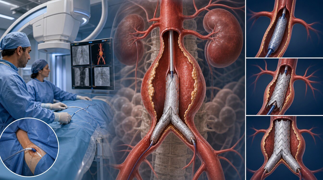
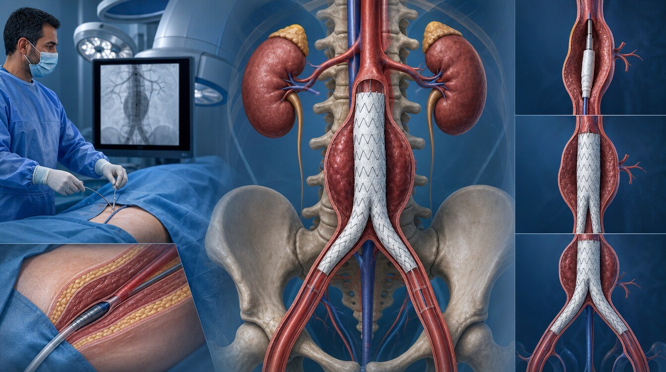
The usual access is the femoral artery. Entry can be through a small surgical cut or through the skin with a needle and guidewire. The stent-graft is a fabric-like covered prosthesis strengthened by a metal scaffold. Once it is in place, the main blood flow passes through it. X-ray images and contrast check position, the relation to important branches and whether there is a leak. The access site may be closed with sutures, a closure device or both.
The frame below is not an operator manual.
- CTA, medicines, kidney function and the anaesthesia plan are reviewed.
- The groin artery is entered; guidewires and catheters are advanced.
- The stent-graft is taken to healthier vessel segments above and below the aneurysm and opened.
- Leak and patency of important branches are checked.
- The access site is closed and leg circulation is watched.
Hospital stay, medicines and recovery
Length of stay depends on whether the procedure was planned or emergency, the access method, any extra intervention and the person’s general state. In the first hours the groins, leg pulses, warmth and colour, blood pressure, urine output, kidney function and signs of bleeding or infection are watched.
After planned EVAR, mobilisation can often be earlier than after open surgery; this is not the same for every person. Heavy lifting, driving, return to work and exercise should follow the discharge plan.
A clinician may advise cholesterol-lowering, blood-pressure or antiplatelet medicines according to cardiovascular risk. Medicines should not be stopped without advice.
Possible complications
EVAR is a major vascular treatment. Risk varies with age, kidney and heart status, whether the aneurysm is an emergency, vessel anatomy and the method used.
Types of endoleak
An endoleak is continued leak of blood into the aneurysm sac around, rather than through, the stent-graft.[5,8]
- Type I: leak from the proximal or distal attachment zone. It can transmit high pressure and is usually treated.
- Type II: back-filling from side branches. It is the most common type. It is often watched if the sac is not growing; a procedure may be considered if it grows.
- Type III: separation of stent-graft parts or a tear in the fabric. It usually needs active treatment.
- Type IV: temporary microscopic leak through the fabric after the procedure. It is uncommon with newer devices and usually settles on its own.
- Type V: sac growth without a clear leak on imaging (endotension). The cause is not always clear.
Type I and III endoleaks are linked to a higher rupture risk. Endoleaks can appear immediately or years later.[5,8]
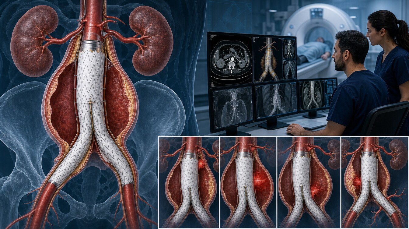
Even if the stent-graft is in place, the sac may not shrink or may grow. Migration is movement of the stent-graft. Separation of parts, kinking or occlusion of a limb can cause sudden leg pain, coldness, paleness or weakness. Loss of internal-iliac flow can impair circulation in the hip muscles.[2]
The access site can bleed, bruise, collect lymph, become infected, tear or impair leg circulation. Contrast used for CTA and during the procedure can affect kidney function, especially if kidney function is already reduced.
Other uncommon but serious complications include embolisation, bowel ischaemia, stent-graft infection, heart attack, arrhythmia, stroke, conversion to open surgery, later rupture of the aneurysm, a further procedure and X-ray or contrast exposure.
Outcomes and evidence
Randomised trials comparing EVAR with open repair have shown that early results and long-term results are not the same, especially in planned treatment. Most of these trials used earlier-generation stent-grafts; today’s devices, imaging, planning and follow-up practice can differ.[4,5,9]
In the UK EVAR-1 trial, 1252 people were randomised to EVAR or open repair with early-generation devices and followed for a mean of 12.7 years. Total and aneurysm-related death looked lower in the EVAR group in the first 6 months; that early survival advantage was lost over time. After eight years, total and aneurysm-related death were lower in the open-repair group; a substantial part of late aneurysm-related death after EVAR was linked to secondary sac rupture. The main message is the importance of lifelong imaging and further procedures when needed.[4]
In OVER, 881 people were followed for up to 14 years. Long-term overall survival was similar after EVAR and open repair; trends favouring EVAR in the first years and open repair in some later periods did not form a statistically conclusive difference. Secondary procedures were more common after EVAR.[5]
Older randomised trials such as DREAM and ACE also showed a balance between early results, durability, further procedures and patient selection. ACE reported that in people at low to moderate surgical risk, open repair could be as safe as EVAR and more durable.[9]
The reading of the evidence is different in emergency ruptured aneurysms. In the IMPROVE randomised trial, an “endovascular-first strategy; open repair if anatomy is not suitable” approach was compared with immediate open repair for suspected rupture. There was no clear, statistically conclusive difference in one-year death, while discharge was faster in the endovascular-strategy group. At three years death was lower in the endovascular-strategy group; at seven years there was no clear mortality difference between the groups. These results show that anatomy, an experienced team and the centre’s facilities are decisive in emergency care.[6]
These rates must not be used for TEVAR, trauma or type B dissection.
Emergency warning symptoms
Suitability and non-suitability
EVAR may be considered when repair is discussed because of diameter, growth or symptoms; CTA shows adequate landing zones for the ends of the stent-graft; proximal neck length, diameter and angulation fit the chosen device IFU; the femoral and iliac arteries will accept the delivery system; and a plan can protect flow in important branches.
Standard EVAR may be less suitable when the neck is very short, highly angulated, irregular or lined with thrombus or calcium; the vessel diameter or shape will not hold the stent-graft safely; the groin and iliac arteries are very narrow, tortuous or heavily calcified; important branches would inevitably be covered; there is an active stent-graft infection; or lifelong follow-up is not possible.
“Not suitable” sometimes applies only to standard EVAR; a more complex endovascular technique or open repair may still be possible.
Lifelong follow-up
EVAR does not remove the aneurysm sac from the body. Position of the stent-graft, attachment of its ends, sac diameter and possible endoleaks can change over time. Follow-up after EVAR should therefore continue for life.[2,8]
Duplex ultrasound, CTA or, in selected cases, MR angiography can be used. The timing of the first check and later intervals follow the early imaging result, anatomical conditions, whether the sac is shrinking and kidney function. In some people with a satisfactory first check, yearly ultrasound may be enough; the vascular team providing follow-up should make that decision.[2,8]
Contact the team without waiting for an appointment if there is new abdominal or loin pain, sac growth on review, a new endoleak on imaging, a sudden circulation problem in a leg, fever, or growing swelling, redness or discharge at the groin access site.
Decision path
- AAA is diagnosed and the clinical state (intact, symptomatic, leaking or ruptured) is separated.
- If there is an emergency finding, an EVAR strategy or open repair is considered according to anatomy.
- If the picture is stable, diameter, growth, symptoms and life expectancy are weighed between surveillance and repair.
- If repair is on the table, CTA is used to review the neck, iliac access and branches against the IFU.
- Standard EVAR, fenestrated or branched EVAR, open repair or surveillance are compared.
- A shared decision is made; if EVAR is chosen, a lifelong imaging plan is set.
Questions for an Aortic Team
- What is the exact site and maximum diameter of my aneurysm? Is it infrarenal or does it extend aorto-iliac?
- How fast has it grown? Is there a symptom that raises the rupture risk now?
- What are the length, diameter and angulation of my aortic neck? Does my anatomy fit the device instructions for use?
- Are my groin and iliac arteries adequate? How will the renal, mesenteric or internal iliac arteries be protected?
- What are the different risks of open repair and EVAR for me?
- When is my first follow-up scan, and which method will be used?
- Which symptoms mean I should go to emergency care?
Academic context of Prof. Dr. Hakan Uçar
Among the sources that could be verified for this package, no confirmed EVAR outcome paper by Prof. Dr. Hakan Uçar was found. No specific device, procedure count or success rate is attributed to him.
Frequently asked questions
No. In EVAR the aneurysm sac is usually left in place. The stent-graft is placed so that blood is directed away from the inside of the sac. The sac may shrink over time; follow-up is still needed for growth or endoleak.
EVAR is not a classic open abdominal operation. Access is usually from the groin arteries. It is still a major vascular procedure, and some people need a surgical cut, extra vessel repair or conversion to open repair.
Not always. General, local or regional anaesthesia may be used. The choice follows the extent of the procedure, urgency and general health.
The time depends on the shape of the aneurysm, the type of stent-graft, how difficult access is and any extra steps. The treating team gives a personal estimate.
Not always. The aim is to reduce or stop blood flow into the sac. Shrinkage can be a favourable finding; no change or growth needs further assessment.
CTA shows the arteries in more detail using contrast and X-rays. Ultrasound does not use radiation and can be enough for some follow-up. The choice follows previous imaging, kidney function and the clinical question.
Kidney disease does not automatically rule EVAR out; contrast and procedural risk are judged more carefully. Less contrast or alternative imaging can be planned.
Recovery time depends on the extent of the procedure, the access method and general health. Activity is usually increased step by step. Heavy lifting, exercise and return to work should follow the personal discharge plan.
Some people are given medicines to manage arterial and cardiovascular risk. The choice is linked more to overall vascular disease than to the aneurysm itself. Medicines should not be started or stopped without a clinician’s advice.
It is an advanced endovascular repair that uses stent-grafts with special openings or side branches so that flow to important arteries such as the renal or mesenteric arteries can continue. Planning is more complex than standard EVAR, and secondary procedures and follow-up remain important.[2]
In some people, if the anatomy is suitable and an experienced emergency vascular team and the needed facilities are available, an endovascular-first strategy can be used. If the anatomy is not suitable, open repair may be needed. A suspected rupture is not decided like planned treatment.[6]
Literature
References
Mazzolai L, Teixidó-Turà G, Lanzi S, et al. 2024 ESC Guidelines for the management of peripheral arterial and aortic diseases. European Heart Journal. 2024;45:3538–3700. DOI: 10.1093/eurheartj/ehae179
Wanhainen A, Van Herzeele I, Bastos Goncalves F, et al. Editor's Choice — European Society for Vascular Surgery (ESVS) 2024 Clinical Practice Guidelines on the Management of Abdominal Aorto-iliac Artery Aneurysms. European Journal of Vascular and Endovascular Surgery. 2024;67:192–331. DOI: 10.1016/j.ejvs.2023.11.002
Chaikof EL, Dalman RL, Eskandari MK, et al. The Society for Vascular Surgery practice guidelines on the care of patients with an abdominal aortic aneurysm. Journal of Vascular Surgery. 2018;67:2–77.e2. DOI: 10.1016/j.jvs.2017.10.044
Patel R, Sweeting MJ, Powell JT, Greenhalgh RM; EVAR trial investigators. Endovascular versus open repair of abdominal aortic aneurysm in 15-years' follow-up of the UK EVAR trial 1. The Lancet. 2016;388:2366–2374. DOI: 10.1016/S0140-6736(16)31135-7
Lederle FA, Kyriakides TC, Stroupe KT, et al. Open versus Endovascular Repair of Abdominal Aortic Aneurysm. New England Journal of Medicine. 2019;380:2126–2135. DOI: 10.1056/NEJMoa1715955
IMPROVE Trial Investigators. Comparative clinical effectiveness of endovascular strategy versus open repair for ruptured abdominal aortic aneurysm: three year results of the IMPROVE randomised trial. BMJ. 2017;359:j4859. DOI: 10.1136/bmj.j4859
US Preventive Services Task Force. Screening for Abdominal Aortic Aneurysm: US Preventive Services Task Force Recommendation Statement. JAMA. 2019;322:2211–2218. DOI: 10.1001/jama.2019.18928
Smith T, Quencer KB. Best Practice Guidelines: Imaging Surveillance After Endovascular Aneurysm Repair. American Journal of Roentgenology. 2020. DOI: NA. PMID: NA.
Becquemin JP, Pillet JC, Lescalie F, et al. A randomized controlled trial of endovascular aneurysm repair versus open surgery for abdominal aortic aneurysms in patients with a low to moderate surgical risk (ACE). Journal of Vascular Surgery. 2011;53:1167–1173. DOI: 10.1016/j.jvs.2010.10.124
This content is for general information only and does not replace personalized medical advice. In emergencies call local emergency services.
Continue