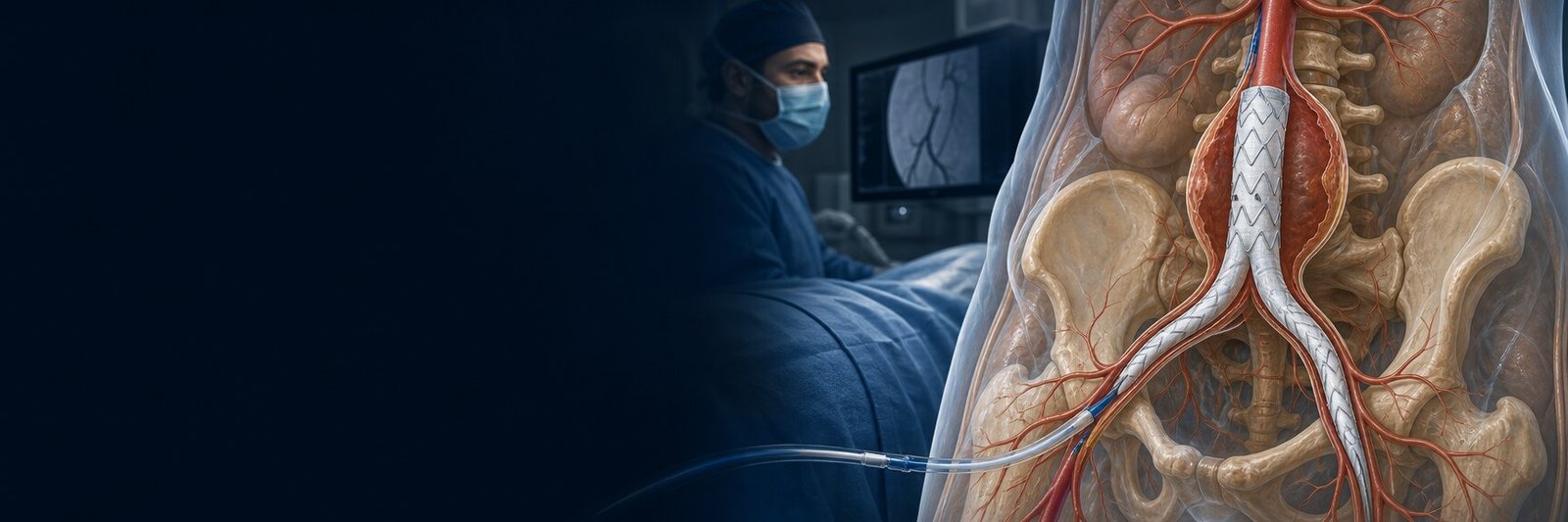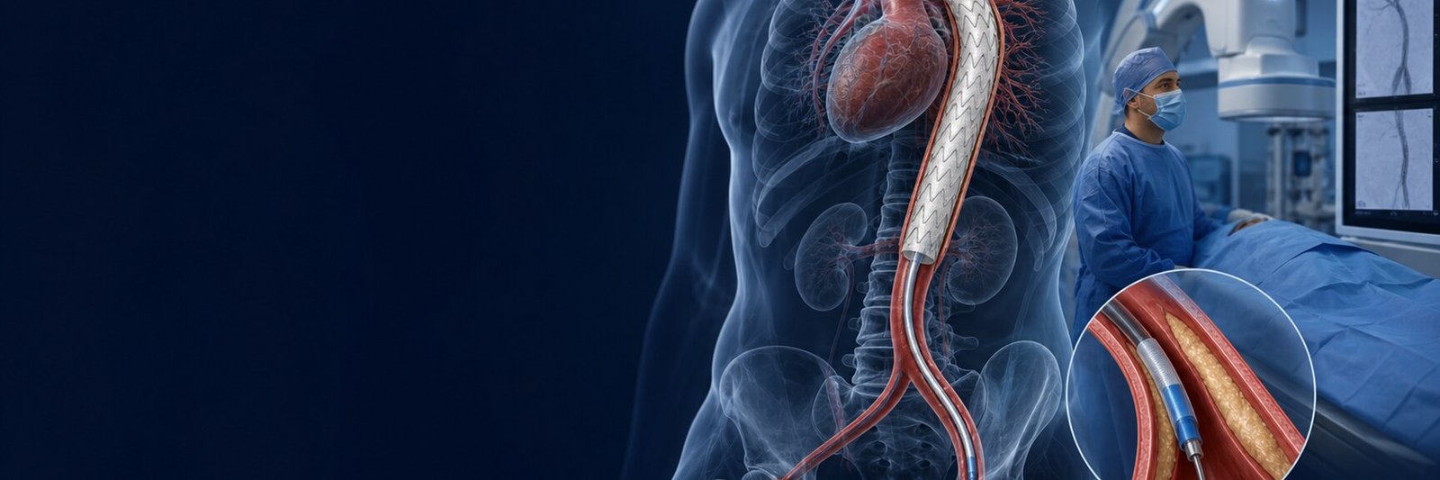
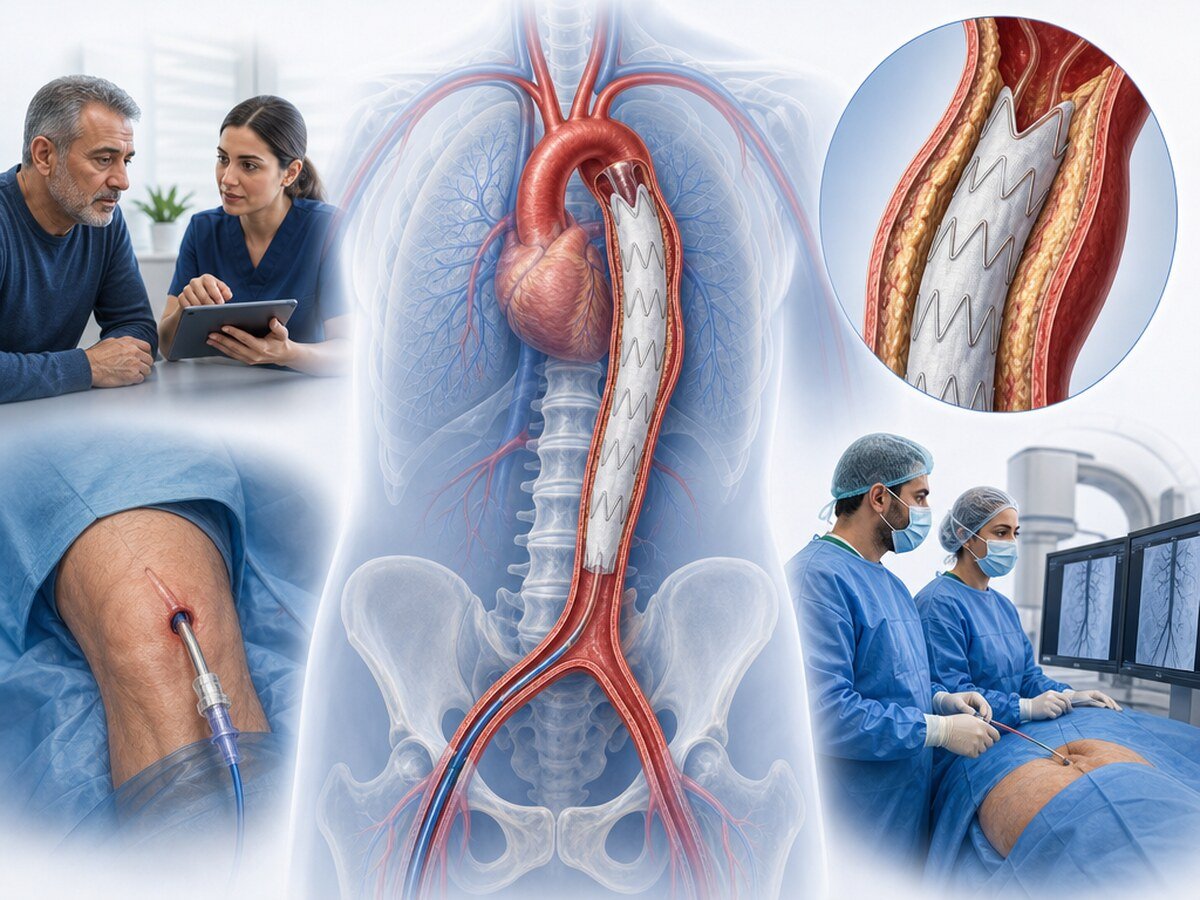
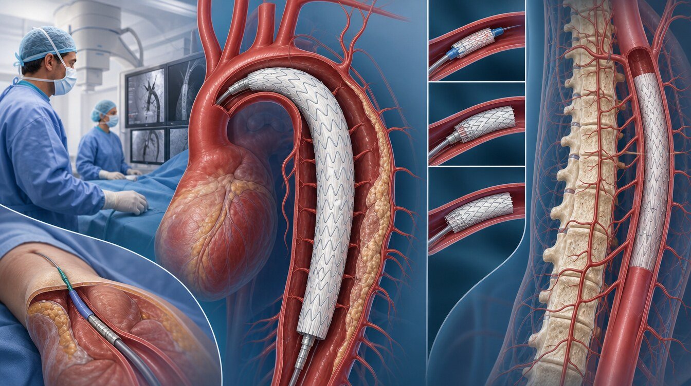
TEVAR (thoracic endovascular aortic repair) treats a diseased segment of the aorta inside the chest with a covered stent-graft placed from inside the arteries rather than by open surgery. The aim is to take blood flow away from the diseased wall, reduce the risk of tearing or further widening and keep aortic continuity.[1–3]
During TEVAR the clinician usually advances a thin catheter and delivery system from the femoral artery in the groin. The stent-graft opens onto healthier aortic segments before and after the diseased area. Those contact zones are called landing zones.
TEVAR is not suitable for every thoracic aortic disease. The type of disease, the segment, vessel diameter, age and general health, the position of aortic branches and whether the anatomy fits the device instructions for use (IFU) are judged together. Guidelines emphasise that the decision should be made by an experienced, multidisciplinary Aortic Team.[1,2]
This article is a child page of aortic aneurysm and endovascular treatment. EVAR is a separate procedure for the abdominal aorta; AAA diameter thresholds, EVAR randomised results and EVAR endoleak rates are not transferred to TEVAR diseases.
In which diseases is TEVAR used?
The five pictures below are different. Results obtained for one disease are not automatically transferred to another.
Degenerative descending thoracic aortic aneurysm
An aneurysm can develop over time in the descending thoracic aorta. Degenerative aneurysm is most often linked to ageing, wall weakness, hypertension and atherosclerosis. The treatment decision is not based on diameter alone; growth rate, pain, shape, wall features and operative risk are also considered.
Surveillance and medical risk reduction can be enough; in some conditions TEVAR or open repair comes into view. In isolated descending thoracic aortic aneurysm, when both TEVAR and open repair are suitable, guidelines support TEVAR for short-term hospital results and recovery burden; the need for long-term follow-up remains.[3] That evidence is not direct outcome evidence for type B dissection, traumatic injury or a penetrating aortic ulcer.
Complicated type B aortic dissection
Aortic dissection is a tear between the layers of the aortic wall, so that blood creates a new channel inside the wall. Type B dissection usually extends beyond the left subclavian artery and does not involve the ascending aorta.
Complicated type B dissection means reduced blood flow to organs, rapid aortic widening, ongoing or uncontrolled pain, bleeding, rupture or circulatory failure. In that picture the usual aim of TEVAR is to close the entry tear that started the dissection and to direct blood into the true lumen. In uncomplicated type B dissection the first approach in most people is blood-pressure and heart-rate control with close imaging; evidence gaps remain about whether early TEVAR prevents chronic complications.[4]
Dissection results must not be combined with aneurysm results.
Penetrating aortic ulcer and intramural haematoma
A penetrating aortic ulcer is deep erosion of the aortic wall by an atherosclerotic plaque. An intramural haematoma is blood collected between the wall layers, usually without a clear flow channel. These two pictures differ from dissection; they can still progress to dissection, aneurysm or rupture.[5]
Asymptomatic, lower-risk cases can be watched with medicines and imaging. Ongoing pain, progression on imaging, signs of rupture or high-risk anatomy can bring TEVAR or another repair into discussion. High-quality comparative evidence in this group is limited.[5]
In one series of symptomatic penetrating ulcer and intramural haematoma, adverse events of intervention, recurrent symptoms, imaging progression, rupture or death were reported in 25 percent of people at 30 days after diagnosis and in about half at one year. Those rates belong only to that group and must not be transferred to other reasons for TEVAR.[5]
Traumatic aortic injury
Traumatic aortic injury is damage to the thoracic aorta, most often after a high-energy road crash or sudden deceleration. The injury can range from a small tear of the inner surface to complete transection.
Some low-grade injuries can be watched with blood-pressure control and serial imaging. In more serious or progressive injuries TEVAR is often considered. There is no randomised controlled trial proving that TEVAR is superior to open surgery; the available evidence comes mainly from retrospective registries, series and observational studies.[6]
In a retrospective study of 382 people from nine trauma centres, among 198 people treated with TEVAR, device malposition was reported in 3.0 percent, endoleak in 2.5 percent, stroke in 1.0 percent and spinal-cord injury resulting in paralysis in 0.5 percent. These data belong to a specific trauma cohort; they must not be generalised to elective aneurysm or dissection.[6]
Ruptured thoracic aortic disease
Rupture is tearing of the aortic wall with leakage of blood and is a life-threatening emergency. If anatomy allows, TEVAR can aim to close the bleeding site quickly. Evidence for TEVAR in rupture is mostly observational and from meta-analyses. Findings in the rupture setting must not be transferred to planned degenerative-aneurysm repair.[3]
Why does the aortic segment matter?
Not every region of the aorta is treated in the same way.[1,2]
- Aortic root: next to the valve and the origins of the coronary arteries. Standard TEVAR usually cannot treat this anatomy.
- Ascending aorta: the first segment leaving the heart. Open surgery or hybrid treatments may be needed.
- Aortic arch: the curved segment where branches to the brain and arms arise. Rerouting of vessels, branched devices or hybrid repair may be needed.
- Descending thoracic aorta: the region most often considered for standard TEVAR; adequate proximal and distal landing zones are still required.
- Thoraco-abdominal aorta: chest and abdominal aorta together. Visceral branches must be protected.
- Inherited connective-tissue disease (Marfan, Loeys–Dietz): how the stent-graft sits and how the aorta behaves over time differ. Open or staged hybrid strategies may be more suitable than standard TEVAR.[1,2]
Who is suitable for TEVAR, and who is not?
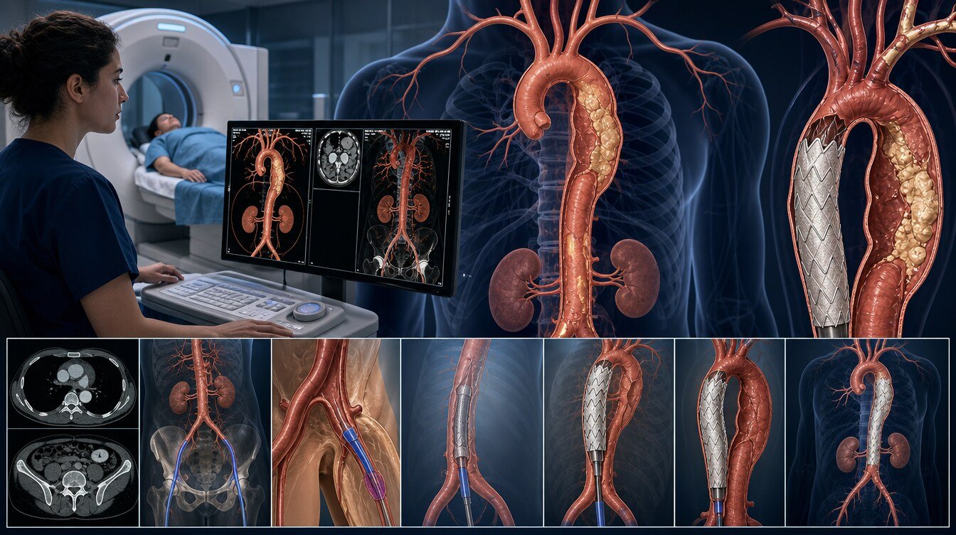
TEVAR can be an option when the disease is in the descending thoracic aorta; there are adequate healthier segments for safe proximal and distal seating; diameter, curves and wall structure fit the device IFU; the groin and pelvic arteries can accept the delivery system; the balance between disease risk and procedural risk is acceptable; and long-term imaging follow-up can be done.[3]
Standard TEVAR may not be suitable when there is no adequate landing zone; the root, ascending aorta or a complex arch is involved; renal or mesenteric arteries would be covered; the femoral–iliac arteries are very small, tortuous, calcified or damaged; there is widespread wall weakness from inherited connective-tissue disease; diameter or shape is outside the IFU; or severe kidney failure prevents contrast.
“Not suitable” does not always mean “cannot be treated”. Open, hybrid, branched or fenestrated endovascular or staged options can still be reviewed.
Imaging and TEVAR planning
CTA is a fast examination that shows the arteries in detail with contrast. MRA uses magnetic resonance. In emergencies CTA is often used because of speed and detailed anatomical planning.[3]
If TEVAR is considered, thin-slice imaging of the whole aorta plus the iliac and femoral arteries is usually needed. Imaging of the neck vessels matters, especially for the left subclavian artery and vertebrobasilar circulation.[3]
The main features measured are: the segment of disease; maximum diameter and how it was measured; growth rate; proximal and distal landing zones; whether the left subclavian artery would need to be covered; vessels to the spinal cord, brain, arm and abdominal organs; femoral–iliac access; important sources of spinal-cord blood supply; and IFU limits.
If the left subclavian artery needs to be covered, some people have surgical or endovascular revascularisation beforehand or in the same session. A systematic review has reported that this revascularisation can reduce the risks of stroke and impaired spinal-cord blood flow; much of the evidence is non-randomised and extra complications can occur at the extra procedure site.[7]
How are the treatment options compared?
Surveillance and medical risk reduction. Imaging at intervals, blood-pressure control, stopping smoking and managing cholesterol and other cardiovascular risk. It can be appropriate for some small, asymptomatic or lower-risk diseases. Surveillance does not remove existing wall injury.
Standard TEVAR. It can result in smaller skin incisions, often a shorter hospital stay and a lower early surgical burden. Contrast, access-site problems, endoleak, device movement and later procedures can still occur. TEVAR is not “surgery-free”; it is a major aortic intervention done from inside the vessel.
Branched, fenestrated or hybrid repair. Special openings or side branches are used for organ arteries; in hybrid repair vessels may first be rerouted surgically. Technical results have been reported in thoraco-abdominal aneurysms, but reintervention and continuous follow-up remain important limits.[8]
Open surgery. It replaces the diseased aortic segment and can be the most reliable or the only suitable option in some anatomies. Opening the chest, a longer recovery, lung problems, kidney injury, bleeding and impaired spinal-cord blood flow are among the risks.
No approach is universally superior. Guideline support is clearer for degenerative descending thoracic aneurysm; there is no randomised trial in traumatic aortic injury; in type B dissection, especially timing, uncertainties remain.[3,4,6] In registries, patient selection, newer devices and centre experience can affect results.
How is the procedure done?
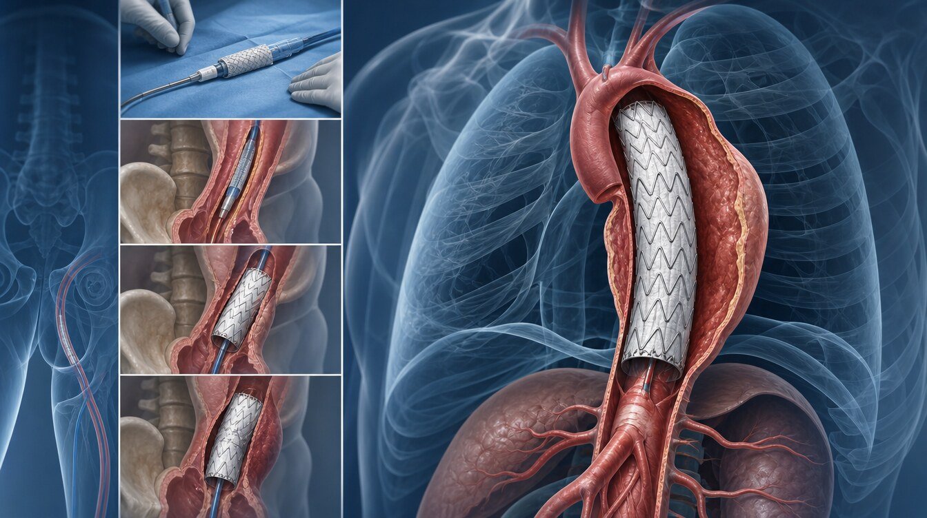
The Aortic Team can bring together cardiac and vascular surgery, interventional radiology, cardiology, anaesthesia and, when needed, neurology, genetics and intensive care. Images can be reviewed with three-dimensional centreline reconstruction.[3]
TEVAR can be done under general anaesthesia or, in some people, regional or local anaesthesia. The femoral artery is entered through a small cut or needle puncture. The stent-graft is opened at the planned site under imaging. Left-subclavian flow is protected if needed.
In people at higher risk of spinal-cord ischaemia, blood-pressure targets, cerebrospinal-fluid drainage or staging the procedure can be considered. The choice follows the person and the centre’s protocol.[3]
The frame below is not an operator manual.
- Disease type, segment, landing zones and spinal-cord risk are planned.
- Anaesthesia and femoral (and if needed extra) access are obtained.
- The stent-graft is advanced to the target site and opened.
- Branches, leak and the neurological state are checked.
- Leg strength, speech and vision are watched in intensive or close care.
Hospital stay, medicines and lifelong follow-up
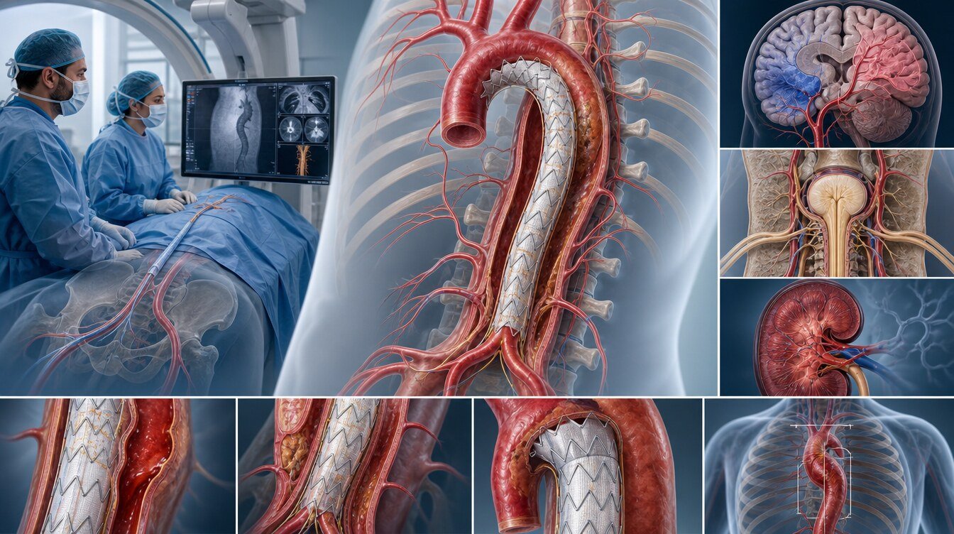
After the procedure, pulse, blood pressure, kidney function, leg circulation and the neurological state are assessed. Length of stay depends on whether the procedure was an emergency, extra interventions and accompanying disease.
Whether an antiplatelet or anticoagulant is needed follows the reason for TEVAR and other diseases; the same medicine is not required in everyone.
Imaging follow-up after TEVAR is lifelong. Endovascular-treatment guidelines recommend contrast CT usually at one and twelve months and then yearly, with closer imaging if there is an endoleak or another problem.[3]
Risks and complications
Risk varies with the type and urgency of the disease, the length of aorta covered, the state of the left subclavian artery, previous procedures and general health.
- Stroke: clot or material reaching the brain arteries, or an effect on brain blood flow.
- Spinal-cord ischaemia and paraplegia: reduced blood flow to the spinal cord; risk can rise with long-segment coverage and previous aortic procedures.
- Retrograde type A dissection: the tear extends towards the heart; uncommon but serious.
- Rupture: internal bleeding and the need for emergency surgery.
- Endoleak: blood leaking around the stent-graft; some are watched, some need a further procedure.
- Graft migration: the stent-graft slides from the planned site.
- Access-vessel injury, embolisation, kidney or contrast injury, mesenteric ischaemia, infection, reintervention, conversion to open surgery.
Emergency warning
A segment-and-disease decision path
- The disease is named: degenerative descending thoracic aneurysm, complicated type B dissection, penetrating ulcer or intramural haematoma, traumatic injury or rupture.
- Urgency is judged. Rupture, organ ischaemia, progressive trauma, uncontrolled pain or rapid widening can need early repair.
- The aortic segment is identified. If disease is limited to the descending thoracic aorta, standard TEVAR may be anatomically possible. Root, ascending, arch or thoraco-abdominal involvement brings open, hybrid or branched-fenestrated approaches into view.
- Landing zones, the left subclavian artery and visceral branches are reviewed.
- Access arteries and the device IFU are checked.
- A plan to reduce spinal-cord and organ ischaemia risk is made.
- Natural disease risk, procedural risk, further procedures and the burden of lifelong follow-up are compared with the person.
Questions for an Aortic Team
- What is the exact name of my aortic disease, and in which segment is it? Is treatment emergency, early or planned?
- What is the aim of TEVAR: to stop bleeding, close a dissection entry or prevent further widening?
- Is the landing zone adequate for standard TEVAR? Will the left subclavian artery be covered?
- Are the femoral and iliac arteries suitable for access? How was IFU fit assessed?
- What are the advantages and disadvantages of open, hybrid, branched-fenestrated repair and surveillance?
- How are my risks of spinal-cord ischaemia, stroke, kidney injury and a further procedure judged?
- When is the first scan, and what is my lifelong follow-up programme?
Limits of the evidence
Evidence on TEVAR is not one type. Guidelines and comparative data exist for degenerative descending thoracic aortic aneurysm, but many studies include people with different risk profiles. TEVAR is an important option in complicated type B dissection, yet consensus is limited on timing in the acute phase, early intervention in uncomplicated dissection and comparison with open surgery in chronic dissection.[4]
High-quality evidence is scarce for penetrating aortic ulcer and intramural haematoma.[5] There is no randomised controlled trial that directly compares TEVAR with open repair for traumatic aortic injury.[6] Rupture data mostly come from emergency registries. In observational studies, people with more suitable anatomy can cluster in the TEVAR group. Device designs and centre experience have changed over time.
Academic context of Prof. Dr. Hakan Uçar
Among the sources that could be verified for this package, no confirmed TEVAR outcome paper by Prof. Dr. Hakan Uçar was found. No specific device, procedure count or success rate is attributed to him. EVAR or abdominal aortic aneurysm papers are not used as TEVAR evidence.
Frequently asked questions
The chest is usually not opened; the procedure is done from inside the arteries. It is still a major aortic intervention, and conversion to open surgery can be needed in some situations.
The choice of anaesthesia follows the extent of the procedure, urgency and general health. General anaesthesia is one of the commonly used options; other methods can be considered in some people.
The time depends on the site of the disease, the length of aorta to be covered, how difficult access is and any extra steps. A guaranteed duration cannot be given in advance.
Pain during the procedure is usually controlled by anaesthesia. Afterwards there can be pain or tenderness in the groin; severe or increasing pain should be told to the care team.
The stent-graft is usually left permanently inside the artery. Removal is considered only in special complications or when a different treatment is needed.
TEVAR aims to reduce blood flow and wall pressure in the diseased segment; it does not mean the whole aorta is “cured”. Disease can progress in other segments, and follow-up is needed.
The same medicine is not needed in every person. Antiplatelet or anticoagulant treatment follows the reason for TEVAR, accompanying heart-rhythm or arterial disease and bleeding risk.
Early, controlled walking is encouraged in most people; the timing depends on the extent of the procedure and the state of the groin arteries. Heavy exercise and lifting should wait for the treating team’s approval.
Return to desk work can be earlier; return to physically demanding work can be later. The personal advice follows urgency, complications and the nature of the job.
Aortic disease and pregnancy together can carry special risk. A pregnancy plan should not be made without review by cardiology, obstetrics, genetics and, when needed, aortic specialists.
MRA can be suitable for some follow-up; in emergencies and for device planning, CTA can be more practical or more detailed. The choice follows kidney function, device features and the clinical question.
Covering this artery can be necessary in some people, but arm, brain and spinal-cord circulation can be affected. Whether revascularisation is needed is therefore judged from the anatomy.[7]
In some people a further procedure is needed because of an endoleak, device movement, new aortic widening or disease in another segment. That chance varies with the type of disease and long-term imaging.
Literature
References
Mazzolai L, Teixidó-Turà G, Lanzi S, et al. 2024 ESC Guidelines for the management of peripheral arterial and aortic diseases. European Heart Journal. 2024;45:3538–3700. DOI: 10.1093/eurheartj/ehae179
Isselbacher EM, Preventza O, Hamilton Black J 3rd, et al. 2022 ACC/AHA Guideline for the Diagnosis and Management of Aortic Disease. Circulation. 2022;146:e334–e482. DOI: 10.1161/CIR.0000000000001106
Upchurch GR Jr, Escobar GA, Azizzadeh A, et al. Society for Vascular Surgery clinical practice guidelines of thoracic endovascular aortic repair for descending thoracic aortic aneurysms. Journal of Vascular Surgery. 2021;73:55S–83S. DOI: 10.1016/j.jvs.2020.05.076
MacGillivray TE, Gleason TG, Patel HJ, et al. The Society of Thoracic Surgeons/American Association for Thoracic Surgery clinical practice guidelines on the management of type B aortic dissection. Journal of Thoracic and Cardiovascular Surgery. 2022. DOI: NA. PMID: NA.
Evangelista A, Czerny M, Nienaber C, et al. Interdisciplinary expert consensus on management of type B intramural haematoma and penetrating aortic ulcer. European Journal of Cardio-Thoracic Surgery. 2015. DOI: NA. PMID: NA.
Scalea TM, Feliciano DV, DuBose JJ, et al. Blunt thoracic aortic injury: multicenter observational outcomes after TEVAR. Observational trauma cohort. Year and DOI: NA. PMID: NA.
Waterford SD, Chou D, Bombien R, et al. Left subclavian arterial coverage and revascularization during thoracic endovascular aortic repair. Systematic review. DOI: NA. PMID: NA.
Oderich GS, Tenorio ER, Mendes BC, et al. Fenestrated and branched endografts for complex aortic aneurysms. Technical series; reintervention and follow-up remain limitations. DOI: NA. PMID: NA.
This content is for general information only and does not replace personalized medical advice. In emergencies call local emergency services.
Continue