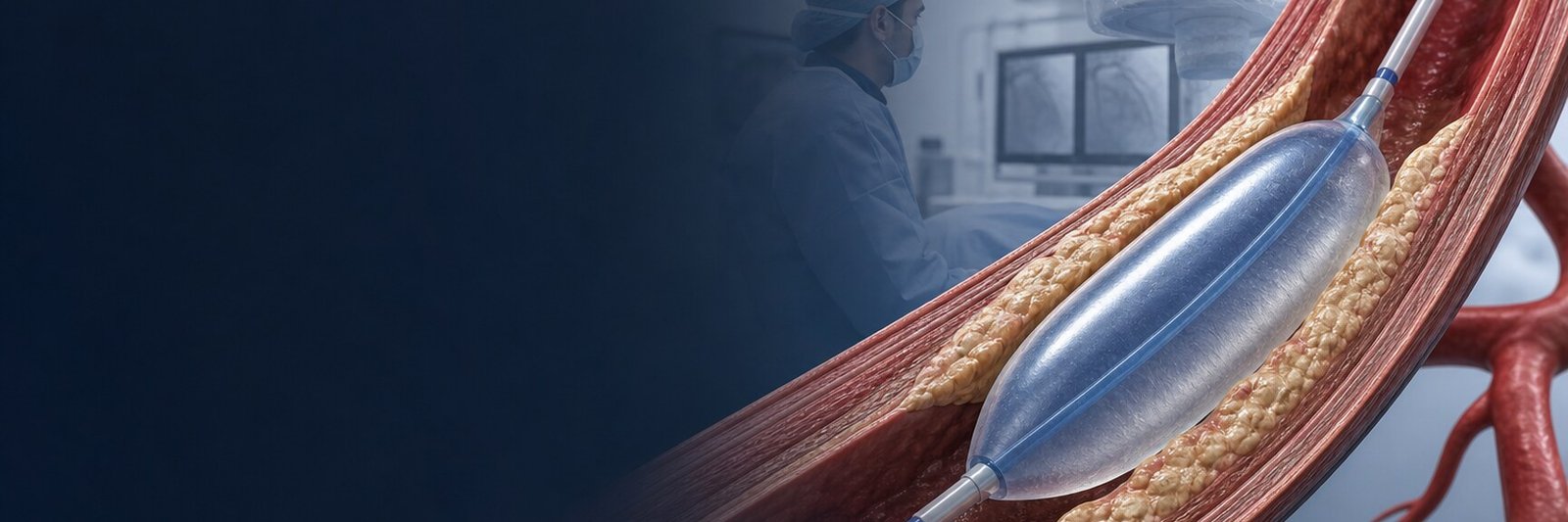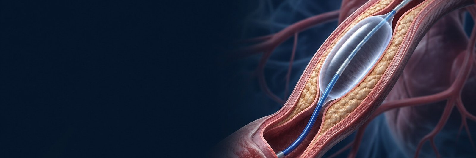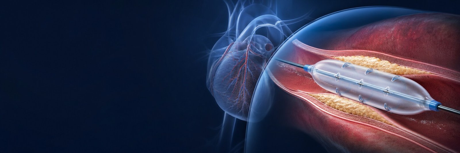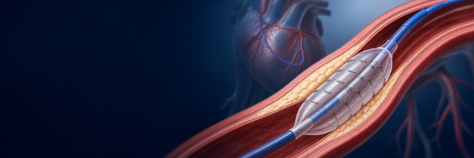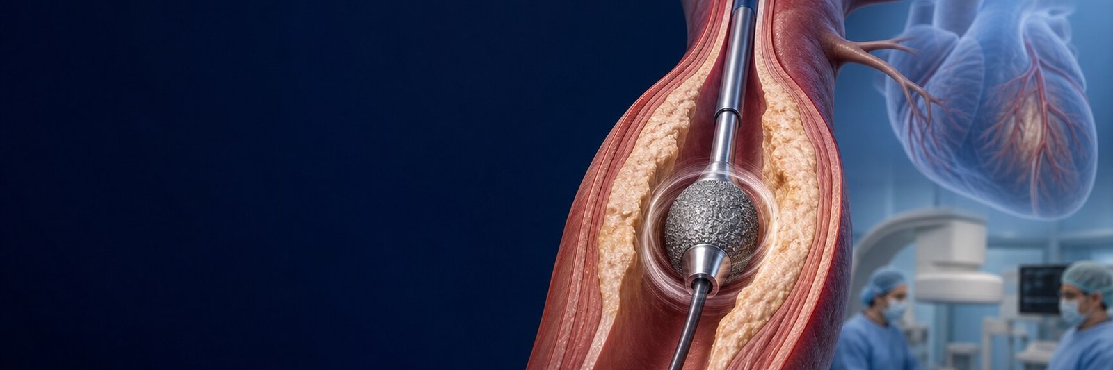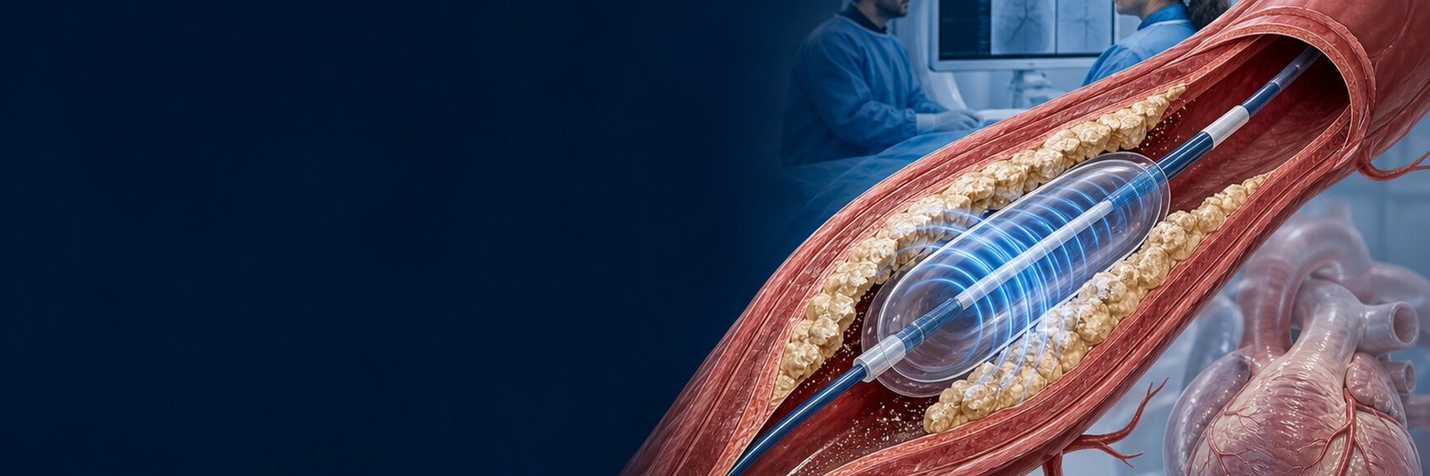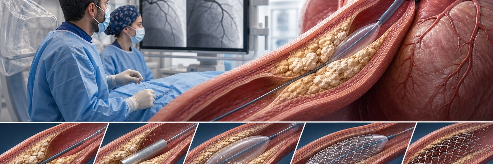
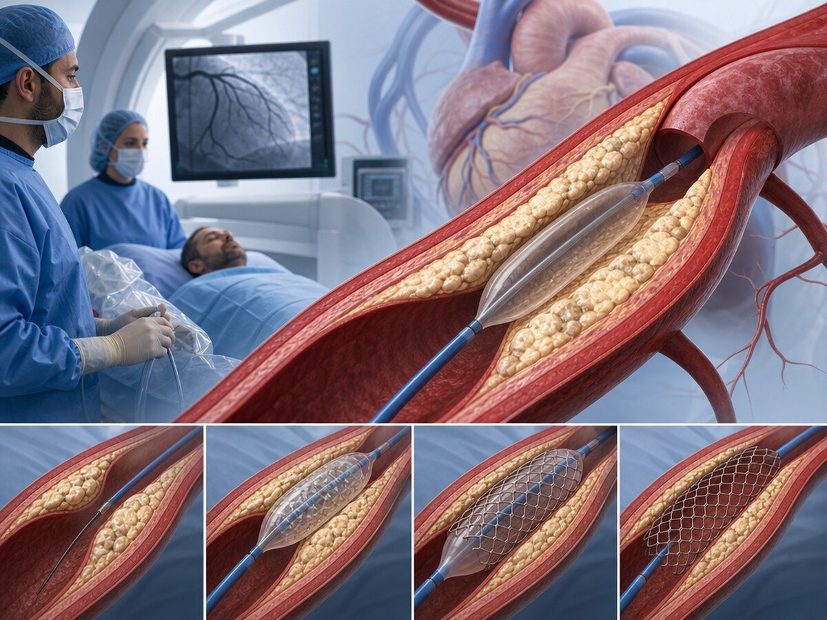
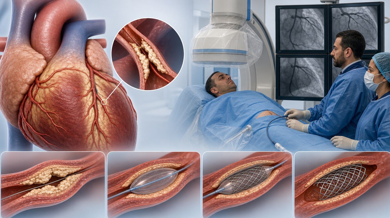
Calcified coronary lesion means a rigid stenotic area caused by substantial calcium within plaque in the wall of a coronary artery. It may reduce arterial flexibility and make balloon expansion, stent delivery or full stent expansion more difficult.[1–4]
Treatment planning therefore asks more than “which device?”. The team assesses whether the stenosis needs treatment, whether calcium is superficial or deep, how far it extends around the vessel, whether balloons or imaging catheters can cross it, and whether a stent is likely to expand adequately.[2–4]
Is a calcified coronary lesion the same as a calcium score?
No. The concepts are related but answer different questions:
- A coronary artery calcium score, usually measured by non-contrast cardiac CT, quantifies total coronary calcium burden to help estimate future cardiovascular risk.
- A calcified coronary lesion describes how calcium at one stenosis may affect PCI and stenting during angiography or intravascular imaging.
A high calcium score does not by itself mean a particular artery needs a stent. Conversely, angiography can reveal a heavily calcified lesion that complicates treatment even when a calcium score was never measured.[2,3]
Why does coronary calcification matter?
Calcium-containing plaque can be rigid and poorly compliant, causing several procedural problems:
- A wire may cross while a balloon or stent cannot reach the lesion.
- A balloon may not fully expand despite high pressure.
- A stent may under-expand or fail to appose fully.
- Dissection or vessel-wall injury may occur at balloon or stent edges.
- Device-specific complications can include slow flow, perforation or device entrapment.
- An under-expanded stent can increase later restenosis or stent-thrombosis risk.[1–5]
This does not mean every calcified lesion needs a special device. Moderate calcium may respond to suitable balloon preparation, whereas heavy or balloon-undilatable calcium may require additional modification.[1–4]
How is the severity of calcification assessed?
Coronary angiography
Angiography may show calcium as radiopaque densities along the artery. Its two-dimensional view can underestimate calcium thickness and circumferential extent.[3,4]
IVUS
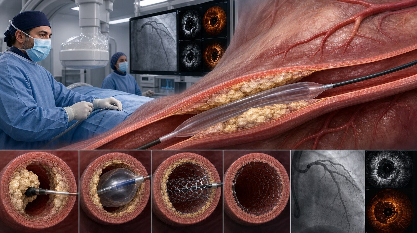
Intravascular ultrasound (IVUS) creates cross-sectional vessel images with sound waves. It helps assess calcium arc, length, nodules, vessel size and stent expansion, but acoustic shadowing limits direct thickness measurement.[3,4]
OCT
Optical coherence tomography (OCT) provides high-resolution light-based imaging and can usually show calcium arc, length and thickness. The Fujino OCT score combines wide arc, length and thickness features to help predict stent under-expansion, but it is not an absolute treatment rule.[3,4,6]
| Method | What does it show? | Main limitation |
|---|---|---|
| Angiography | Stenosis location, flow and visible calcium | May underestimate depth and circumferential extent |
| IVUS | Vessel size, calcium arc/length, nodules and stent expansion | Limited direct measurement of calcium thickness |
| OCT | Calcium arc, length, thickness, fractures and stent apposition | Requires additional contrast and catheter passage |
Why may an ordinary balloon fail to expand?
Soft, compliant plaque deforms more readily during inflation, whereas dense calcium can resist balloon pressure. Expansion at both ends while the centre remains constrained is sometimes called a dog-bone appearance. Uncontrolled pressure escalation can increase vessel-wall injury instead of preparing the lesion.[3,4]
Failure to expand is therefore not simply a “use more pressure” problem. Vessel and balloon size, calcium shape and depth are reassessed, and another balloon or calcium-modification method may be selected.[3,4]
Which methods can be used for calcified coronary lesions?
Non-compliant (NC) balloon
NC balloons change diameter only slightly as pressure rises and allow controlled dilatation. They may be used for initial preparation in moderate calcium or post-dilatation after stenting. If the balloon cannot cross or fully expand, another method may be needed.[3,4]
Super high-pressure balloon
Double-layer super high-pressure balloons may be considered when a lesion can be crossed but not expanded with a standard NC balloon. Permitted pressure depends on that device's instructions and cannot be generalised. Off-axis or inappropriate inflation can increase vessel injury.[3,4,9]
Scoring and cutting balloons
A scoring balloon concentrates force along external scoring elements; a cutting balloon uses small blades to create controlled linear plaque modification. They are not the same device. Crossability, vessel size, tortuosity and lesion location influence selection.[3,4]
Rotational atherectomy
Rotational atherectomy advances a small diamond-coated burr over a dedicated wire to modify superficial hard tissue into microscopic particles, improving lesion crossability and dilatability. It is not open surgery and does not scrape all calcium out of the artery.[1,3,4]
It may be considered in very tight balloon- or imaging-uncrossable lesions and superficial or nodular calcium. Specific risks include slow flow, vessel injury, transient rhythm disturbance and burr entrapment.[1,3,4]
Orbital atherectomy
An eccentrically rotating diamond-coated crown enlarges its orbit to modify calcified plaque. It is neither the same device nor the same evidence category as rotational atherectomy. Vessel size, tortuosity and crossability matter.[1,3,4,12]
Intravascular lithotripsy (IVL)
Coronary IVL uses pressure waves from a special balloon inflated within the lesion to create fractures in vessel-wall calcium. The balloon and catheter are removed and no lithotripsy device remains in the body.[1,3,4,10,11,15]
The IVL balloon must cross the lesion. It may be considered for deep or circumferential calcium, larger vessels or inadequate expansion after other preparation. It is not the same procedure as external kidney-stone lithotripsy.[3,4]
Excimer laser and combined methods
Excimer laser may be considered for selected fibrocalcific lesions that standard equipment cannot cross or for particular mechanisms of stent under-expansion. Evidence is more limited than for some other methods.[3,4,11]
One method may not be enough. Atherectomy can first create a pathway and IVL can then modify deeper calcium. Such combined or sequential strategies are not routine and are used according to planned or emerging procedural needs.[3,4]
How is the treatment method selected?
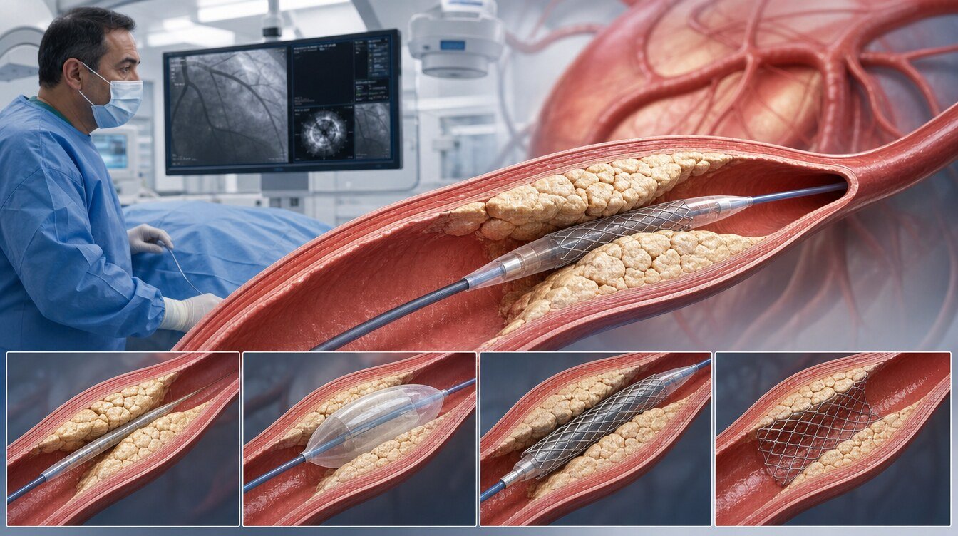
Decisions usually combine these questions:
- Can a balloon cross? IVL or a larger balloon cannot be used if the lesion is uncrossable; a lower-profile or ablation-based method may be needed first.
- Does the balloon cross but fail to expand? Super high-pressure balloons, atherotomy balloons, atherectomy or IVL may be considered according to morphology.
- Is calcium superficial or deep? Imaging helps match mechanism to location, although much of this matching is based on consensus and mechanics rather than clinical-outcome RCTs.[3,4]
- Is there a calcified nodule? Nodules can affect expansion and long-term outcome and may need imaging and specific preparation.
- Is the vessel tortuous, ostial, bifurcated or left main? Side-branch wire protection, deliverability and vessel size change selection.
- Is a stent already present? Calcium-related under-expansion differs from new tissue growth; IVUS/OCT helps identify the mechanism.[5]
- Is this an acute myocardial infarction? Thrombus, flow, haemodynamics and urgency alter planning.
What does the scientific evidence show?
Many studies measure procedural or imaging endpoints such as device crossing, balloon expansion, calcium fracture, residual stenosis and minimum stent area. These matter, but do not by themselves prove fewer deaths, myocardial infarctions or repeat procedures.[3,4,16]
- PREPARE-CALC found greater strategy success with upfront rotational atherectomy than modified balloons in severe calcium, without a clear two-year clinical-endpoint advantage.[7,8]
- ISAR-CALC found that a super high-pressure balloon could achieve a larger final lumen and less residual stenosis than a scoring balloon after inadequate NC-balloon expansion; it was not powered for long-term death or infarction.[9]
- In ROTA.shock, IVL was non-inferior to rotational atherectomy for minimum stent area, with similar expansion.[10]
- ROLLER COASTR-EPIC22 compared rotational atherectomy, IVL and excimer laser. IVL was non-inferior to rotational atherectomy for OCT-measured expansion; laser missed the specified intention-to-treat margin. It was not designed to prove clinical-event superiority.[11]
- ECLIPSE enrolled 2,005 patients eligible for either strategy; routine orbital atherectomy did not reduce one-year target-vessel failure or significantly increase minimum stent area versus balloon angioplasty.[12]
- CALIPSO showed improved imaging measures of stent placement and expansion with an OCT-guided assessment and preparation algorithm versus angiography guidance. Long-term clinical-event reduction remains unproved.[13]
- BALI showed a lower one-year composite procedural/clinical endpoint when IVL was added to conventional preparation; the difference was driven largely by lower OCT residual area stenosis, while safety endpoints did not differ.[14]
- DISRUPT CAD I–IV support feasibility and procedural success of IVL in selected severe calcium. They were single-arm performance-goal studies, not active-comparator randomised trials, and do not prove superiority over other devices.[15]
A 2025 systematic review of 31 randomised trials found no preparation method with demonstrated superiority for all-cause death or serious adverse events; most trials lacked power for these clinical outcomes.[16]
How is the procedure generally performed?
Calcium modification is performed in a catheter laboratory equipped for coronary angiography and PCI:
- Clinical decision: Symptoms, ischaemia, anatomy and medical treatment determine whether PCI is needed.
- Vascular access: The radial or femoral artery is prepared under local anaesthesia.
- Angiography: Lesion location, flow and visible calcium are assessed.
- Guidewire: A fine wire is advanced beyond the stenosis.
- Imaging: IVUS or OCT assesses vessel size and calcium morphology when feasible.
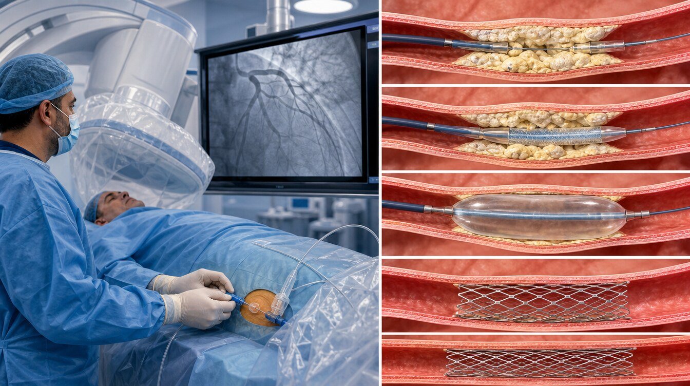
- Lesion preparation: A selected balloon, atherectomy, IVL or laser method is used and may be changed or combined.
- Result check: Balloon expansion, lumen gain and any calcium fracture are assessed.
- Stent/other treatment: Most de novo lesions receive an appropriately expanded stent; selected cases may use another strategy.
- Final imaging: Flow, stent expansion, edge dissection and vessel integrity are checked.
- Monitoring: Access site, rhythm, blood pressure, chest pain and general condition are observed.
Duration and equipment depend on lesion number, location, crossability and the result obtained. Most patients remain awake. Brief chest pressure can occur during balloon inflation and should be reported.[17]
Is a stent always placed after calcium modification?
In PCI for a new calcified lesion, modification is usually performed to allow appropriate stent delivery and expansion. Balloon, atherectomy and IVL devices do not remain; most patients receive a drug-eluting stent.[1–4]
Not every stenosis requires PCI or stenting. Selected lesions may use implant-free strategies such as a drug-coated balloon, according to clinical setting, vessel size, post-preparation result, dissection, flow and re-occlusion risk.
Calcium-related under-expansion of a previous stent is a different problem. IVUS/OCT assessment and expansion of the existing stent are prioritised before adding another stent.[5]
What are the risks?
Risk depends on technique, age, kidney function, ventricular function, acute infarction, anatomy and urgency. Possible complications include:[1–5,17]
- Access-site bruising, bleeding or vascular injury,
- Contrast allergy or kidney impairment,
- Coronary dissection, abrupt closure or perforation,
- Slow flow or no-reflow,
- Transient rhythm or conduction disturbance,
- Device entrapment, balloon damage or wire problems,
- Myocardial infarction, urgent additional intervention or rarely emergency surgery,
- Stent under-expansion, restenosis or stent thrombosis,
- Rare but serious stroke or death.
A complication rate from one study does not describe an individual's risk because lesions, devices, operator experience and event definitions differ.[3,4,16]
Before and after the procedure
What is considered before the procedure?
- Report all medicines, especially antiplatelets, anticoagulants and diabetes medicines.
- Report known contrast, medicine or latex allergies.
- Share kidney disease, dialysis, bleeding history and previous coronary procedures.
- Follow the treating centre's fasting and fluid instructions.
- Do not stop or add medicines without medical advice.
- Bring prior angiograms, stent cards and imaging records when available.[17]
What should be considered after the procedure?
Observation depends on urgency, access route, complexity, kidney function and complications. Same-day discharge may be possible for selected uncomplicated patients but is not universal after extensive calcium modification.[17–19]
After discharge:
- Take prescribed antiplatelet and cardiac medicines as directed.
- Watch for increasing swelling, redness, discharge or bleeding at the access site.
- Follow personal instructions for lifting, driving, work and exercise.
- Attend follow-up and cardiac rehabilitation when advised.
- Smoking cessation and cholesterol, blood pressure and diabetes control remain part of treatment.
Fixed timings in foreign patient leaflets are not universal rules; the personal discharge document and treating team's advice take priority.[17–19]
Which symptoms require emergency help?
Do not wait for routine review if any of these develop:
- New severe chest pain or pain that does not settle with rest,
- Marked breathlessness, fainting or cold sweat,
- Access-site bleeding that does not stop with pressure or rapidly enlarging swelling,
- Coldness, pallor, bluish colour or numbness in the hand or foot on the treated side,
- New speech difficulty, facial droop or one-sided weakness.
Call emergency services for these symptoms. Contact the treating centre promptly for fever or increasing access-site pain, redness or discharge.[17–19]
Frequently asked questions
Current medicines do not dissolve established calcified plaque during a procedure. Treating cholesterol, blood pressure and diabetes remains important to reduce future events and slow disease progression.
A stent is placed in most PCI-treated de novo calcified lesions, but the need depends on the clinical setting, vessel appearance after preparation and the selected strategy.
The access site is numbed with local anaesthetic. Brief chest pressure or pain may occur during balloon inflation and should be reported to the team.[17]
Yes. Adequate preparation and stent expansion aim to reduce risk but cannot eliminate restenosis. Medicines and risk-factor control remain important.
Assessment and decision
Treating a calcified coronary lesion is a broader clinical decision process than choosing one device. The need for PCI is established first; calcium distribution, lesion crossability and intravascular imaging are then assessed. Balloon, atherectomy, IVL or a combined strategy may be reconsidered throughout the procedure.
Literature
References
Lawton JS, Tamis-Holland JE, Bangalore S, et al. 2021 ACC/AHA/SCAI Guideline for Coronary Artery Revascularization. Circulation. 2022;145:e18-e114. DOI: 10.1161/CIR.0000000000001038.
Vrints C, Andreotti F, Koskinas KC, et al. 2024 ESC Guidelines for chronic coronary syndromes. Eur Heart J. 2024;45:3415-3537. DOI: 10.1093/eurheartj/ehae177.
Barbato E, Shlofmitz E, Milkas A, et al. Management strategies for heavily calcified coronary stenoses: EAPCI consensus. Eur Heart J. 2023;44:4340-4356. DOI: 10.1093/eurheartj/ehad342.
Riley RF, Patel MP, Abbott JD, et al. SCAI Expert Consensus Statement on Calcified Coronary Lesions. JSCAI. 2024;3:101259. DOI: 10.1016/j.jscai.2023.101259.
Klein LW, Nathan S, Maehara A, et al. SCAI Expert Consensus on In-Stent Restenosis and Stent Thrombosis. JSCAI. 2023;2:100971. DOI: 10.1016/j.jscai.2023.100971.
Fujino A, Mintz GS, Matsumura M, et al. OCT-based calcium score to predict stent underexpansion. EuroIntervention. 2018;13:e2182-e2189. DOI: 10.4244/EIJ-D-17-00962.
Abdel-Wahab M, Toelg R, Byrne RA, et al. PREPARE-CALC Trial. Circ Cardiovasc Interv. 2018;11:e007415. DOI: 10.1161/CIRCINTERVENTIONS.118.007415.
Allali A, Richardt G, Toelg R, et al. Two-year outcomes of PREPARE-CALC. EuroIntervention. 2023. DOI: 10.4244/EIJ-D-22-00677.
Rheude T, Rai H, Richardt G, et al. ISAR-CALC randomised trial. EuroIntervention. 2021;17:481-488. DOI: 10.4244/EIJ-D-20-01000.
Blachutzik F, Meier S, Weissner M, et al. ROTA.shock trial. Catheter Cardiovasc Interv. 2023;102:823-833. DOI: 10.1002/ccd.30815.
Jurado-Román A, Gómez-Menchero A, et al. ROLLER COASTR-EPIC22 Trial. JACC Cardiovasc Interv. 2025;18:606-618. DOI: 10.1016/j.jcin.2024.11.012.
Kirtane AJ, Généreux P, et al. ECLIPSE randomised trial. Lancet. 2025;405:1240-1251. DOI: 10.1016/S0140-6736(25)00450-700450-7).
Amabile N, Rangé G, Landolff Q, et al. CALIPSO Randomized Clinical Trial. JAMA Cardiol. 2025;10:666-675. DOI: 10.1001/jamacardio.2025.0741.
Kristensen AT, Faltermeier P, Kamp C, et al. BALI Trial. JACC Cardiovasc Interv. 2026;19. DOI: 10.1016/j.jcin.2025.09.028.
Kereiakes DJ, Di Mario C, Riley RF, et al. Pooled Analysis of Disrupt CAD Studies. JACC Cardiovasc Interv. 2021;14:1337-1348. DOI: 10.1016/j.jcin.2021.04.015.
Kristensen AT, Faltermeier P, Kamp C, et al. PCI for Calcified Lesions: Systematic Review and Meta-analyses. Interventional Cardiology. 2025. DOI: 10.15420/icr.2025.10.
American Heart Association. Cardiac Catheterization. Reviewed 21 February 2025. Accessed 25 August 2026.
NHS. Coronary angioplasty and stent insertion — Recovery. Accessed 25 August 2026.
MedlinePlus. Cardiac catheterization — discharge. Accessed 25 August 2026.
Continue