
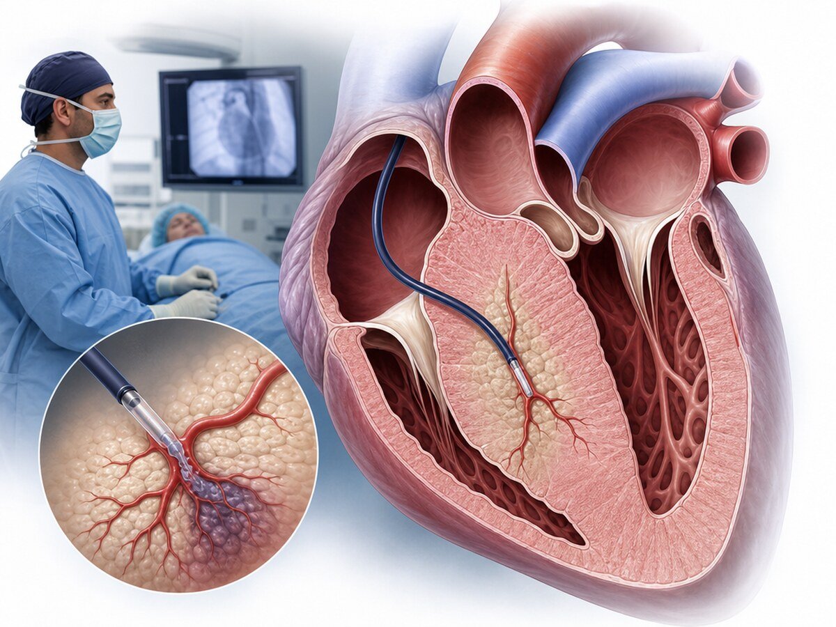
Alcohol septal ablation (ASA) is a catheter-based septal reduction treatment considered in selected people with obstructive hypertrophic cardiomyopathy (HCM) who still have marked symptoms despite medical therapy and who have meaningful dynamic obstruction of the left ventricular outflow tract (LVOT). During the procedure, a controlled amount of ethanol is given into a suitable small coronary branch that supplies the limited part of the thickened septum contributing to the obstruction. The aim is that the planned, limited myocardial injury and later remodelling will reduce the left-ventricular outflow pressure difference.[1,2,5]
What are hypertrophic cardiomyopathy and LVOT obstruction?
Hypertrophic cardiomyopathy (HCM) is a heart-muscle disease in which the myocardium thickens in a way that cannot be fully explained by another loading cause, and which often has genetic features. The disease is not only septal thickness. Relaxation of the heart, mitral-valve motion, intracardiac pressures, rhythm and, in some people, the risk of sudden cardiac death are assessed separately.[1,2]
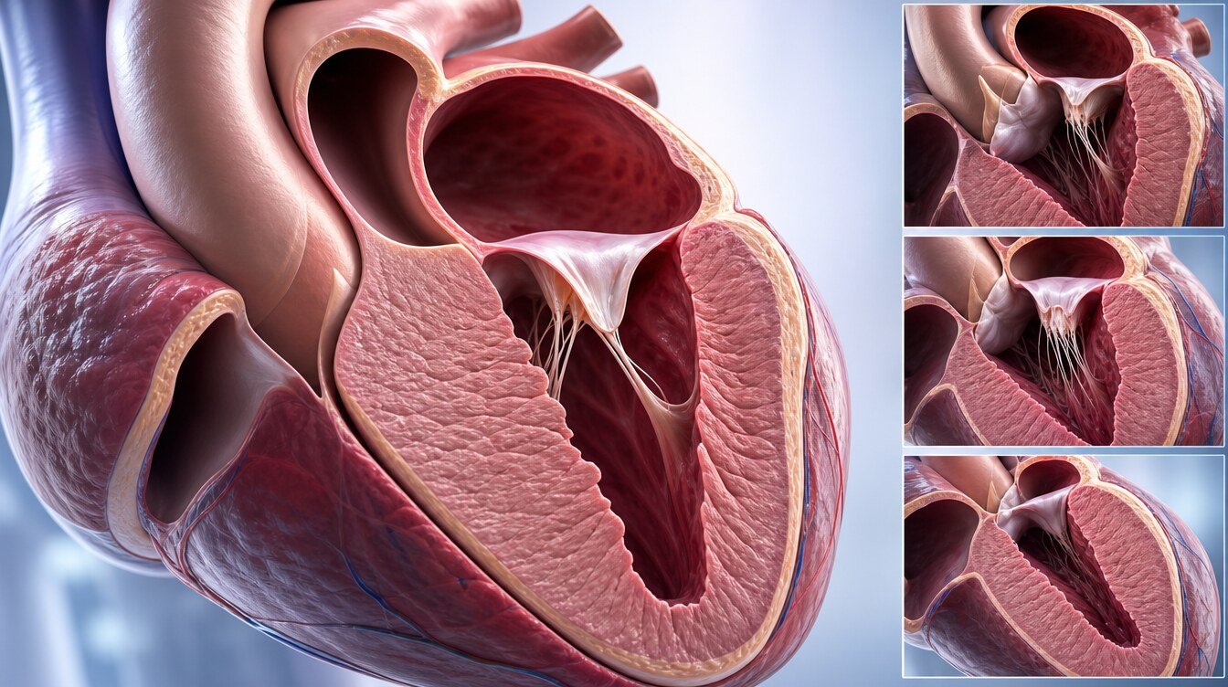
Thickened basal septum together with systolic anterior motion (SAM) of the mitral valve can dynamically narrow the path of blood from the left ventricle into the aorta. This region is called the left ventricular outflow tract, and the measured pressure difference is the LVOT gradient. The gradient may be low at rest and rise with a Valsalva manoeuvre, standing or exercise. A single resting echocardiogram may therefore not show the full clinical effect of obstruction.[1,2]
LVOT obstruction may be related to breathlessness on effort, chest pain, dizziness, fainting, palpitations or reduced exercise capacity. The same symptoms can also come from a rhythm disturbance, coronary disease, heart failure, lung disease or another cause. The treatment decision rests on the relationship between the symptoms and the obstruction, not on the gradient number alone.
How does alcohol septal ablation work?
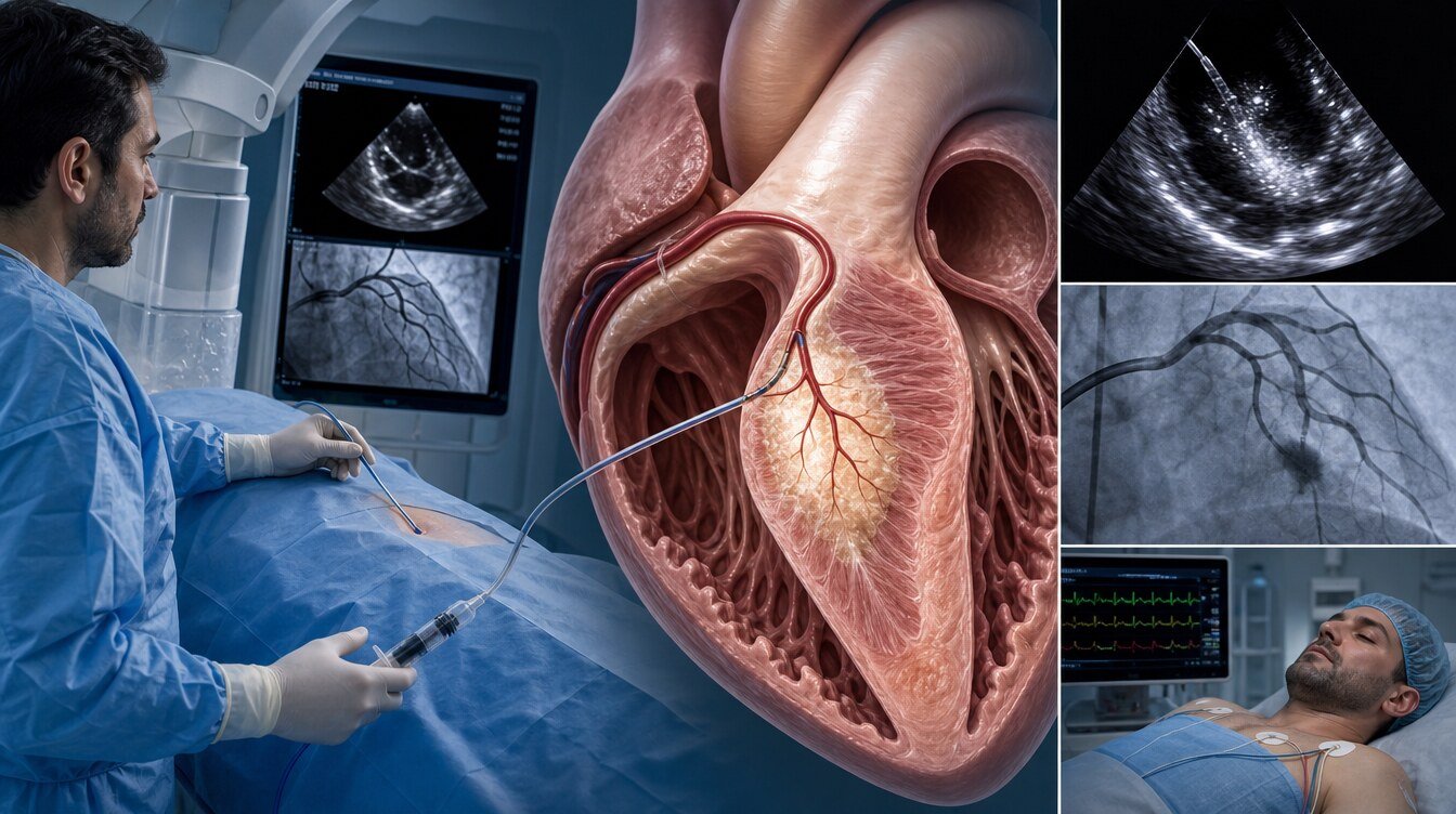
During the procedure, coronary angiography shows the small septal perforator branches that supply the septum. A guidewire and balloon catheter are advanced into a branch that looks suitable. While the balloon temporarily occludes the vessel, echocardiography contrast given through the catheter shows the myocardial territory that vessel supplies. If the target area matches the basal septum that contributes to obstruction, and there is no spread to the right ventricle, a papillary muscle or another unwanted region, a controlled amount of ethanol is given.[5]
Ethanol creates a planned and limited infarction in the target septal tissue. Reduced contraction is expected at first; over the following weeks and months, healing, scarring and remodelling are expected to reduce septal mass. The mechanism is therefore not “dissolving the heart muscle”; it is creating controlled injury in the target tissue. Chest pain, a rise in cardiac enzymes and ECG changes can therefore be part of the expected biological effect, while the team also watches for off-target injury and complications.[1,5]
How does it differ from rhythm ablation?
Alcohol septal ablation and catheter ablation for heart rhythm disorders are not the same procedure.
- Rhythm ablation targets the electrical focus, pathway or circuit that starts or sustains an arrhythmia; radiofrequency, cryoenergy or pulsed field ablation may be used.
- Alcohol septal ablation aims to reduce septal thickening and the related dynamic LVOT obstruction in obstructive HCM; ethanol is given into a suitable septal coronary branch.
ASA is not performed to treat atrial fibrillation or another arrhythmia. If a person with HCM has a rhythm disturbance, rhythm and sudden-death risk are assessed separately; ASA does not replace those assessments or ICD treatment when that is needed.[1,2]
In whom can it be considered?
Current guidelines recommend considering septal reduction, in experienced comprehensive HCM centres, for people with obstructive HCM whose daily life is still clearly limited by symptoms despite optimal medical therapy.[1,2] The following components are usually expected together:
- Confirmation of the HCM diagnosis and of the mechanism of dynamic obstruction
- Marked effort limitation or other clinical symptoms related to obstruction that continue despite medical therapy
- A clinically meaningful LVOT gradient, most often 50 mmHg or higher, at rest or with physiological provocation
- A basal septal target that ASA can reach and a suitable septal perforator coronary anatomy
- It being realistic not to expect mitral-valve, papillary-muscle or other structural problems to be corrected by ASA alone
- Discussion of the benefits and limits of ASA, surgical septal myectomy and medical options in a shared decision process[1,2]
A 50 mmHg threshold is not, by itself, an indication for the procedure. Routine septal reduction is not recommended in an asymptomatic person only to lower one measurement or to prolong life. Guidelines state that there is not enough evidence to support septal reduction solely to improve survival.[1]

In whom may it not be suitable?
ASA may not be suitable, or surgery may come more strongly into view, in the following situations:
- Non-obstructive HCM or a low gradient that has not been shown to relate to the symptoms
- Early procedure assessment before medical therapy has been arranged
- Absence of a suitable target septal perforator vessel
- Contrast showing that the target vessel also supplies unwanted myocardial regions
- A septum that is too thick, or a gradient that is too high, for a sufficient effect to be expected
- Marked mitral-valve disease, an abnormal papillary muscle, an elongated mitral leaflet, a subaortic membrane, severe coronary disease or other heart disease that needs surgical correction in the same session
- A high risk of permanent heart block because of pre-existing conduction disease
- Pregnancy, active infection, an uncontrolled bleeding risk, or another clinical situation that makes contrast or coronary intervention unacceptable
No item is an automatic absolute ban. Suitability is decided individually by the HCM team.[1,2]
How do medical therapy, a myosin inhibitor, ASA and myectomy differ?
In most people with obstructive HCM the first approach is to adjust medicines that affect heart rate and contraction, accompanying diseases and lifestyle triggers. Current guidelines describe options such as beta blockers, suitable non-dihydropyridine calcium-channel blockers, disopyramide in selected situations, or a cardiac myosin inhibitor according to access and suitability.[1,2]
When septal reduction comes into view because marked symptoms continue despite medicines, there are two main approaches:
Surgical septal myectomy
The surgeon removes thickened septal tissue directly. The same session can also correct additional anatomical problems such as mitral-valve, papillary-muscle, coronary-artery or valve disease. In experienced centres it can be used across a wide anatomical range, and the fall in gradient is often more marked. It does, however, require sternotomy, cardiopulmonary bypass and a surgical recovery.[1]
Alcohol septal ablation
It is done through a catheter, does not require sternotomy, and the hospital stay may be shorter. If advanced age, frailty or serious accompanying diseases make surgical risk unacceptably high, if the coronary anatomy is suitable and if the procedure can be done in an experienced HCM centre, it may become the preferred invasive strategy.[1] Permanent pacemaker need and the need for repeat septal reduction may be more frequent than after myectomy; effect may be more limited when the septum is very thick, the gradient is very high or there are additional structural problems.[1,3,7,8]
Is ASA or myectomy more effective?
There is no single answer for every person. There is no randomised trial comparing surgical septal myectomy with ASA; most available data come from observational comparisons of people who differ in age, disease burden and anatomy. People treated with ASA are older and have more accompanying disease in most series. Statistical adjustment cannot remove every unmeasured difference.[1,3,7,8]
Guidelines and current observational data share the following:
- Both methods can reduce symptoms and the LVOT gradient in appropriately selected people.[1–4]
- Myectomy may reduce the gradient more and the need for a repeat procedure is lower.[1,7,8]
- Permanent pacemaker need is higher after ASA.[1,6–8]
- ASA may avoid sternotomy and allow a shorter hospital stay.[1]
- Long-term survival comparisons are conflicting. Some analyses report similar results, while some large-centre data or long-follow-up subgroups have shown a difference in favour of myectomy; patient selection and centre effects prevent a firm causal interpretation.[1,3,7,8]
Age alone therefore does not choose the method. Anatomical suitability, surgical risk, accompanying correctable heart disease, conduction-system risk, the chance of a repeat procedure and the person’s informed preference are considered together.
What assessments are done before the procedure?
Planning usually includes:
- Review of the HCM diagnosis, family history and possible genetic context
- Documentation of the type of symptoms, their relation to effort and their effect on daily life
- Echocardiography at rest and with suitable provocation such as Valsalva, standing or exercise
- Detailed assessment of the septum, SAM, the mitral valve, papillary muscles and subvalvular structures
- Cardiac magnetic resonance to examine the distribution of hypertrophy, fibrosis and alternative diagnoses
- Assessment of coronary artery disease and septal perforator anatomy
- Recording of the ECG, rhythm monitoring and any existing right/left bundle-branch block or AV conduction disease
- Assessment of sudden cardiac death risk and ICD need independently of ASA
- Planning of kidney function, bleeding risk, contrast allergy, and anticoagulant or antiplatelet medicines in use
- Comparison of ASA, myectomy and medical options in the HCM team[1,2,5,6]
Medicines, especially blood thinners, should not be stopped on the person’s own decision. The written procedure plan varies with the centre and with personal risks.
How is alcohol septal ablation performed?
Details can vary with centre protocol and anatomy, but the general steps are:
- Vascular access is obtained; the need for sedation with local anaesthesia is decided.
- A temporary pacemaker catheter may be placed because of the chance of slowing or complete block.
- The coronary arteries are usually reached from a wrist or groin artery.
- Coronary angiography identifies the septal perforator branches.
- A guidewire and a short balloon catheter are advanced into a suitable small branch; the balloon is inflated to occlude the branch temporarily.
- Echocardiography contrast is given through the balloon to show the targeted septal myocardial area.
- If contrast distribution is not suitable, alcohol is not given into that branch; another vessel is assessed or the procedure may be stopped.
- If the target is confirmed, a controlled, limited amount of ethanol is given slowly.
- The gradient, ECG, rhythm, coronary flow and clinical state are reassessed.
- The access site is closed; the person is watched closely for rhythm and conduction disturbances.[1,5]
Starting the procedure does not guarantee that alcohol will be given. If a suitable target vessel or a safe contrast distribution cannot be found, not completing the procedure can be the correct safety decision.
What may be felt during the procedure?
A brief burning or pressure may be felt at the access site. During the planned myocardial injury created by ethanol there may be chest pain, tightness, nausea or a change in rhythm. Pain and circulation are watched by the team and treated as appropriate. Absolute statements such as “it is completely painless” or “nothing is felt” are not accurate.
What are the risks and possible complications?
ASA is an invasive coronary procedure and the risk is not zero. The most important specific risk is high-grade AV block or complete heart block, because the heart’s electrical conduction system is close to the target septum. Temporary block may recover; some people need a permanent pacemaker. In a large risk-model study, findings related to complete block included older age, pre-procedure left bundle-branch block and certain conduction changes during or after the procedure; that model does not replace individual clinical judgement.[6]
Other possible complications include:
- New right or left bundle-branch block, PR prolongation or other conduction changes
- Ventricular or supraventricular rhythm disturbances
- Alcohol reaching myocardium outside the target and an unwanted infarction
- Coronary spasm, dissection, clot or vessel injury
- Bleeding, haematoma, pseudoaneurysm or a more serious vessel problem at the access site
- Fluid around the heart and, rarely, tamponade
- Ventricular septal defect
- An unwanted change in mitral regurgitation
- Stroke or transient ischaemic attack
- Contrast-related kidney injury or an allergic reaction
- Infection
- Need for emergency surgery or another intervention
- Death, very rarely[1,2]
The risk list is not a personal risk calculation. Age, septal thickness, coronary anatomy, existing conduction disease, kidney function, accompanying heart diseases and centre experience change the risk.
Hospital stay and recovery after the procedure
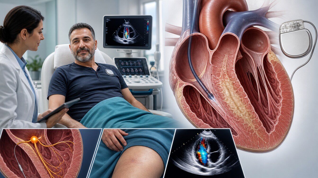
After the procedure the person is usually watched on a rhythm monitor. Conduction disturbance can appear at once or later; the observation period may therefore be longer than after a simple coronary angiogram. A temporary pacemaker may be kept for a defined time according to clinical and ECG findings.[1,6]
Chest discomfort, tiredness, tenderness and bruising at the access site can occur. Discharge is planned from variables such as a stable rhythm, no remaining need for a temporary pacemaker, control of pain and the access site, and kidney function. Written, personal instructions should be followed for driving, return to work, heavy lifting and exercise.
Continuing some medicines does not mean that the procedure has failed. HCM is a disease that needs lifelong follow-up; ASA does not remove the genetic basis, all of the hypertrophy or the arrhythmia risk.
When is the result assessed?
An early fall in the LVOT gradient may be seen in the catheter laboratory; the final effect is not judged from the end-of-procedure measurement alone. Healing and remodelling in the target area develop over weeks to months. Symptoms, examination, ECG and rest/provocation echocardiography are repeated at planned intervals.[1,2]
If symptoms do not improve enough, the reason may be persistent LVOT obstruction, a wrong or incomplete target, mitral–papillary anatomy, diastolic dysfunction, a rhythm disturbance, coronary or lung disease, or another problem. A repeat procedure is not decided from the gradient number alone.
Can repeat ASA or myectomy be needed?
Yes. After ASA, residual or recurrent obstruction and symptoms may lead to repeat ASA or surgical myectomy. Observational comparisons have reported a need for repeat septal reduction more often after ASA than after myectomy.[1,7,8] Surgery can still be possible after a first ASA; previous infarction and conduction changes can, however, affect later planning.
When is urgent help needed after the procedure?
Urgent medical help should be sought if any of the following develops:
- New, severe or unrelieved chest pain
- Marked breathlessness at rest
- Fainting or feeling about to faint
- A very slow, very fast or prolonged palpitation
- Sudden speech disturbance, weakness of the face, arm or leg, or a change in vision
- Bleeding from the access site that does not stop, or rapidly enlarging swelling
- High fever, shivering or signs of infection at the access site
- A marked fall in urine output
- A change in consciousness, cold sweating or a clear decline in general condition
Academic context of Prof. Dr. Hakan Uçar
The 2020 prospective observational study by Prof. Dr. Hakan Uçar and colleagues examined associations between galectin-3 level, clinical findings and the predicted HCM sudden-cardiac-death risk score in 107 people with HCM.[9] The study is an academic example of phenotype and risk assessment in HCM; it is not an alcohol septal ablation outcome, personal procedural experience, a claim of superiority or proof of success. The ASA decision should rest on current guidelines, detailed imaging and a multidisciplinary HCM assessment.[1,2]
Frequently asked questions
It is a catheter procedure that delivers a controlled amount of ethanol into a small coronary branch supplying a selected septal region that contributes to obstruction in obstructive HCM, aiming to create planned, limited myocardial injury and later remodelling.
No. Alcohol is not given into the heart cavity or into the myocardium as a whole; it is applied in a controlled way into a suitable small coronary branch that supplies the target septal tissue.
No. A planned and limited infarction is created in the target tissue; later remodelling is intended to reduce the obstruction.
No. Rhythm ablation targets an electrical arrhythmia focus or circuit; ASA targets septal tissue and LVOT obstruction in obstructive HCM.
Complete heart block can occur in some people and a permanent pacemaker may be needed. Risk is assessed from the pre-procedure ECG and from conduction changes that develop during or after the procedure.[6]
No. ASA aims to reduce obstruction and symptoms; it does not remove the genetic basis of the disease, all of the myocardial thickening or the rhythm risk.
Not automatically. Medicines are adjusted by a clinician according to symptoms, gradient, rhythm, blood pressure and accompanying diseases; a person should not stop a medicine on their own.
In selected people, yes. Scar and conduction changes from a previous ASA can affect surgical planning, so an experienced HCM team is needed.
Literature
References
Ommen SR, Ho CY, Asif IM, et al. 2024 AHA/ACC/AMSSM/HRS/PACES/SCMR Guideline for the Management of Hypertrophic Cardiomyopathy. Circulation. 2024;149:e1239–e1311. DOI: 10.1161/CIR.0000000000001250
Arbelo E, Protonotarios A, Gimeno JR, et al. 2023 ESC Guidelines for the management of cardiomyopathies. European Heart Journal. 2023;44:3503–3626. DOI: 10.1093/eurheartj/ehad194
Cui H, Schaff HV, Wang S, et al. Survival Following Alcohol Septal Ablation or Septal Myectomy for Patients With Obstructive Hypertrophic Cardiomyopathy. Journal of the American College of Cardiology. 2022;79:1647–1655. DOI: 10.1016/j.jacc.2022.02.032
Maurizi N, Antiochos P, Owens A, et al. Long-Term Outcomes After Septal Reduction Therapies in Obstructive Hypertrophic Cardiomyopathy: Insights From the SHARE Registry. Circulation. 2024;150:1377–1390. DOI: 10.1161/CIRCULATIONAHA.124.069378
Nagueh SF, Lakkis NM, He ZX, et al. Role of Myocardial Contrast Echocardiography During Nonsurgical Septal Reduction Therapy for Hypertrophic Obstructive Cardiomyopathy. Journal of the American College of Cardiology. 1998;32:225–229. DOI: 10.1016/S0735-1097(98)00220-4
Karimianpour A, Heizer J, Leaphart D, et al. Predicting Complete Heart Block After Alcohol Septal Ablation for Hypertrophic Cardiomyopathy Using a Risk Stratification Model and Clinical Tool. Catheterization and Cardiovascular Interventions. 2021;98:393–400. DOI: 10.1002/ccd.29478
Bytyçi I, Nistri S, Mörner S, Henein MY. Alcohol Septal Ablation versus Septal Myectomy Treatment of Obstructive Hypertrophic Cardiomyopathy: A Systematic Review and Meta-Analysis. Journal of Clinical Medicine. 2020;9:3062. DOI: 10.3390/jcm9103062
Yokoyama Y, Shimoda T, Shimada YJ, et al. Alcohol Septal Ablation versus Surgical Septal Myectomy of Obstructive Hypertrophic Cardiomyopathy: Systematic Review and Meta-Analysis. European Journal of Cardio-Thoracic Surgery. 2023;63:ezad043. DOI: 10.1093/ejcts/ezad043
Uçar H, Özyılmaz S, Satılmışoğlu MH, Pusuroğlu H. The Importance of Galectin 3 for Risk Stratification and Prognosis in Hypertrophic Cardiomyopathy. EURAS Journal of Health. 2020;1(1):21–35. DOI: 10.17932/EJOH.2020.022/ejoh_v01i1002
This content is for general information only and does not replace personalized medical advice. In emergencies call local emergency services.
Continue