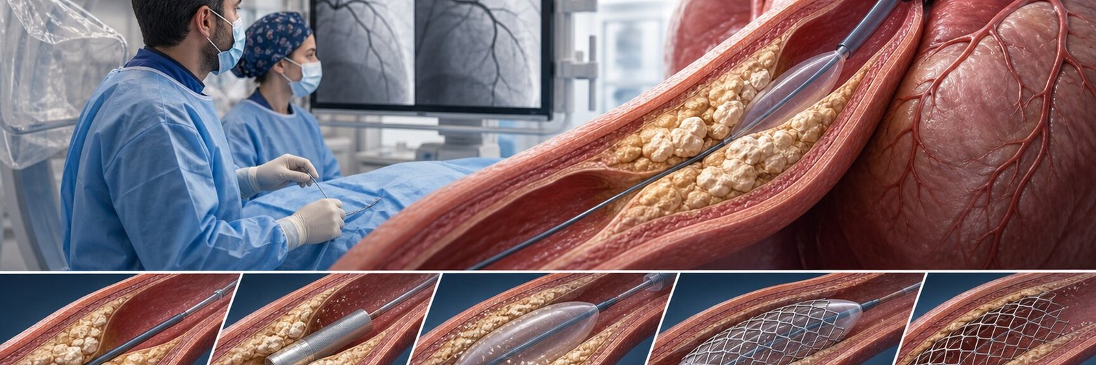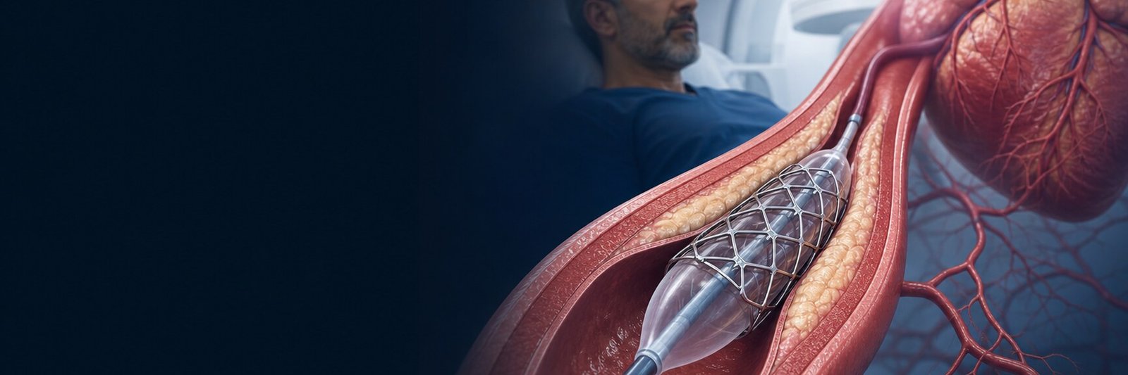
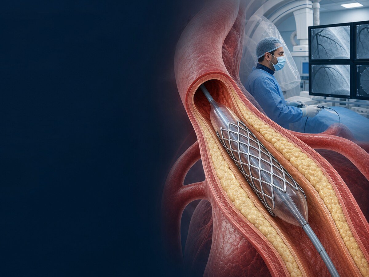
A coronary stent is a small, fine metal-mesh tube left inside a coronary artery after an appropriate narrowing or blockage has been opened. Stent placement is part of a treatment called percutaneous coronary intervention (PCI).
A stent mechanically supports the vessel but does not remove atherosclerosis from the body. Prescribed antiplatelet therapy, cholesterol and blood-pressure control, smoking cessation, activity and nutrition therefore remain integral to treatment.[1–5]
Are coronary stenting and angiography the same procedure?
No. Coronary angiography is a diagnostic procedure that images the arteries. A stent is a treatment applied, when appropriate, to a lesion found on angiography.
After angiography, the path may be medication and lifestyle therapy alone, PCI with balloon and stent, or assessment for coronary bypass surgery. A decision may be made in the same session; complex cases may require image review, additional tests and joint cardiology–cardiac surgery assessment.
How does a stent work?
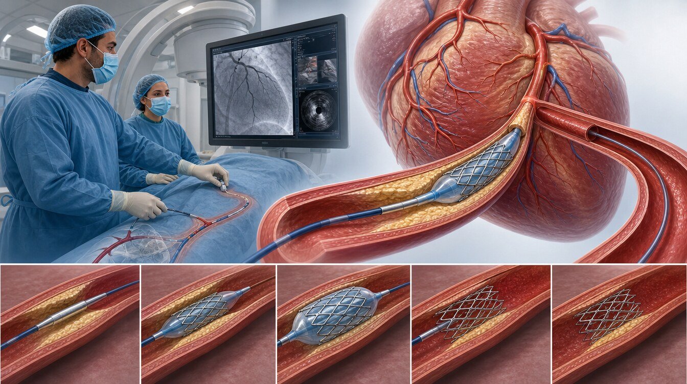
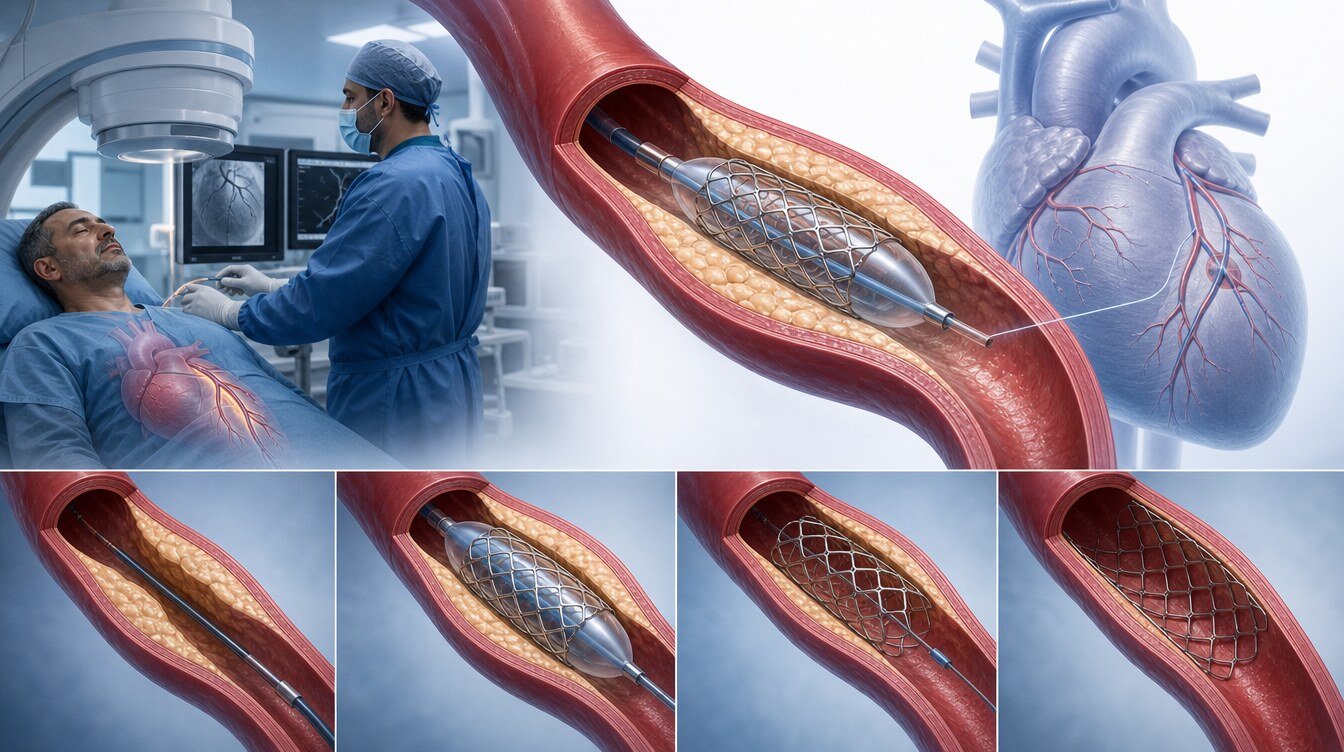
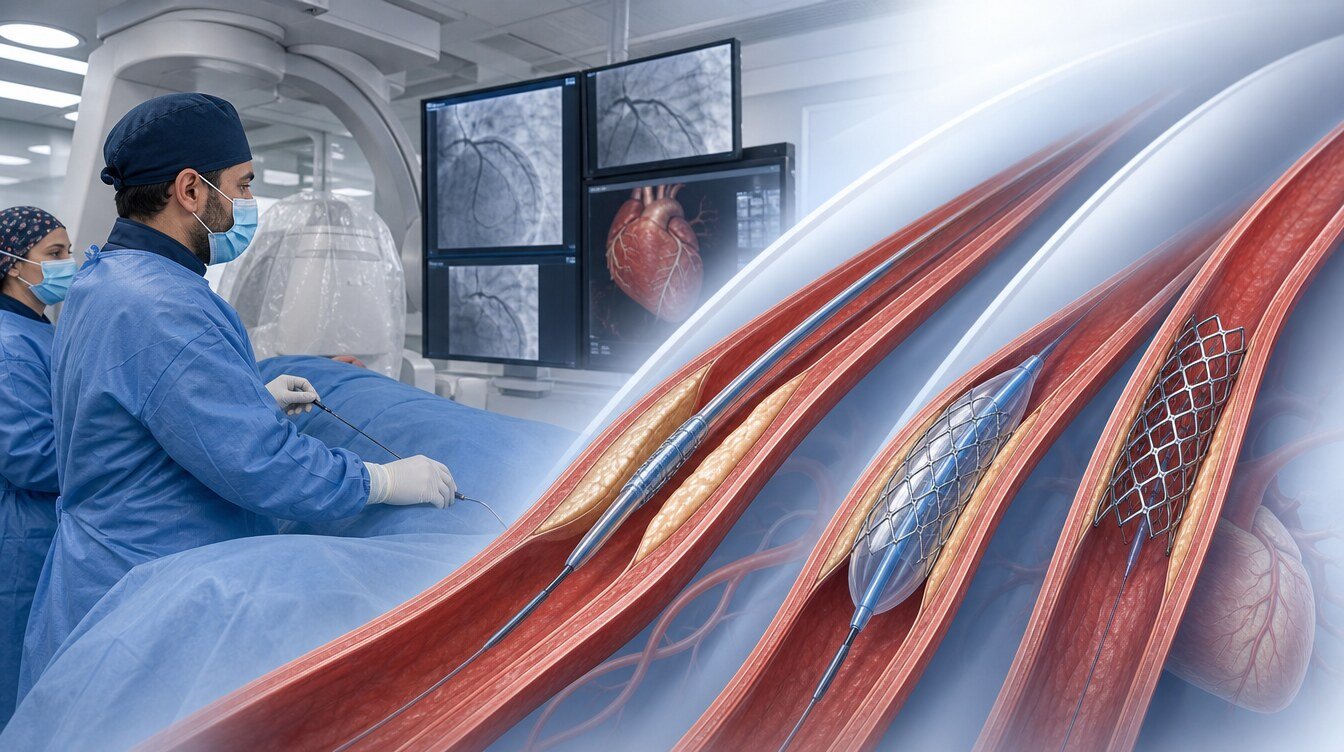
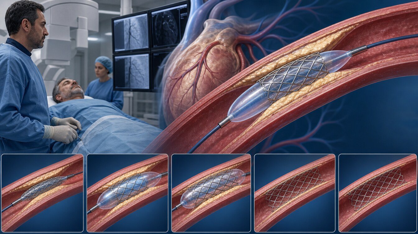
The stent is crimped onto a deflated balloon and carried to the narrowing on a catheter. Inflation expands the stent and reduces the obstruction created by plaque. The balloon and catheter are removed while the stent remains against the vessel wall.
Most current procedures use drug-eluting stents (DES). Medication on their surface aims to reduce excessive tissue growth and recurrent narrowing. Bare-metal stents are historically important, but modern DES are preferred in most current settings.[3,5]
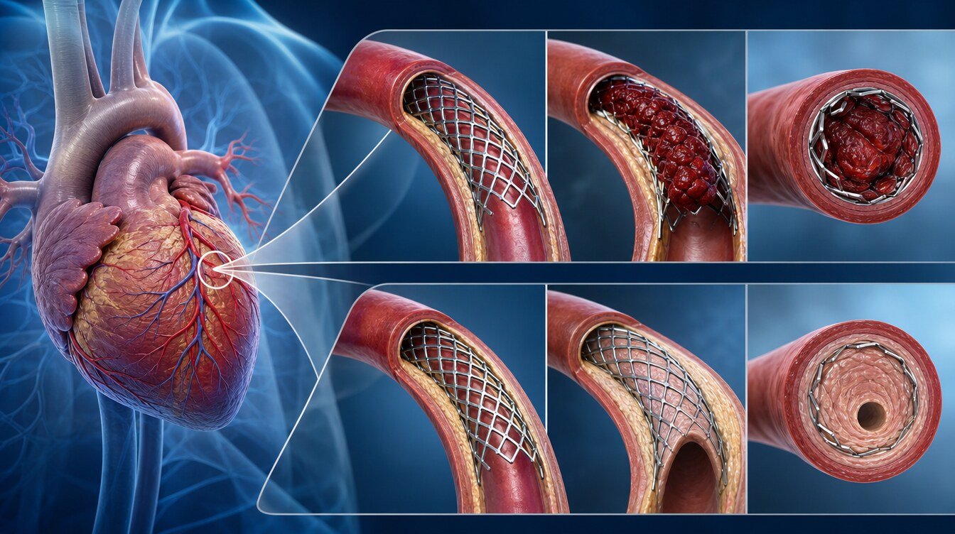
The metal scaffold does not dissolve; tissue grows over it as the inner vessel surface heals. Continuous use of prescribed antiplatelet therapy is particularly important during early healing to prevent clot formation.[1,3,4]
Who needs a coronary stent?
Heart attack and acute coronary syndrome
During a heart attack, a clot may suddenly block a coronary artery or severely reduce flow. In an appropriate patient, emergency angiography and PCI restore blood flow. The aim is not merely symptom relief but protection of jeopardized heart muscle.[1,2]
The 2025 ACC/AHA acute coronary syndrome guideline recommends radial access when suitable and intravascular imaging for complex lesions. With significant multivessel disease, timing and method depend on shock, anatomical complexity and comorbidities.[1]
Stable or chronic coronary disease
In stable disease, the presence of narrowing alone is insufficient. Persistent symptoms despite medication, genuine flow limitation, PCI suitability, left-main or multivessel disease, diabetes, heart function, bypass options, bleeding risk and ability to take long-term medicines are assessed together.
COURAGE and ISCHEMIA showed that routinely adding an invasive strategy to contemporary medication and lifestyle care did not reduce death or major ischaemic events for every stable patient outside specific high-risk groups.[6,7] PCI can improve angina in appropriately selected symptomatic patients. ORBITA and ORBITA-2 also show that symptom benefit depends on selection and background treatment.[9,10]
In FAME 2, PCI for lesions confirmed by FFR to limit blood flow reduced the five-year composite of death, myocardial infarction or urgent revascularization, largely through fewer urgent procedures; mortality did not differ.[8]
How is the need for a stent determined?
Angiography shows anatomy, but the image alone may be insufficient for intermediate lesions.
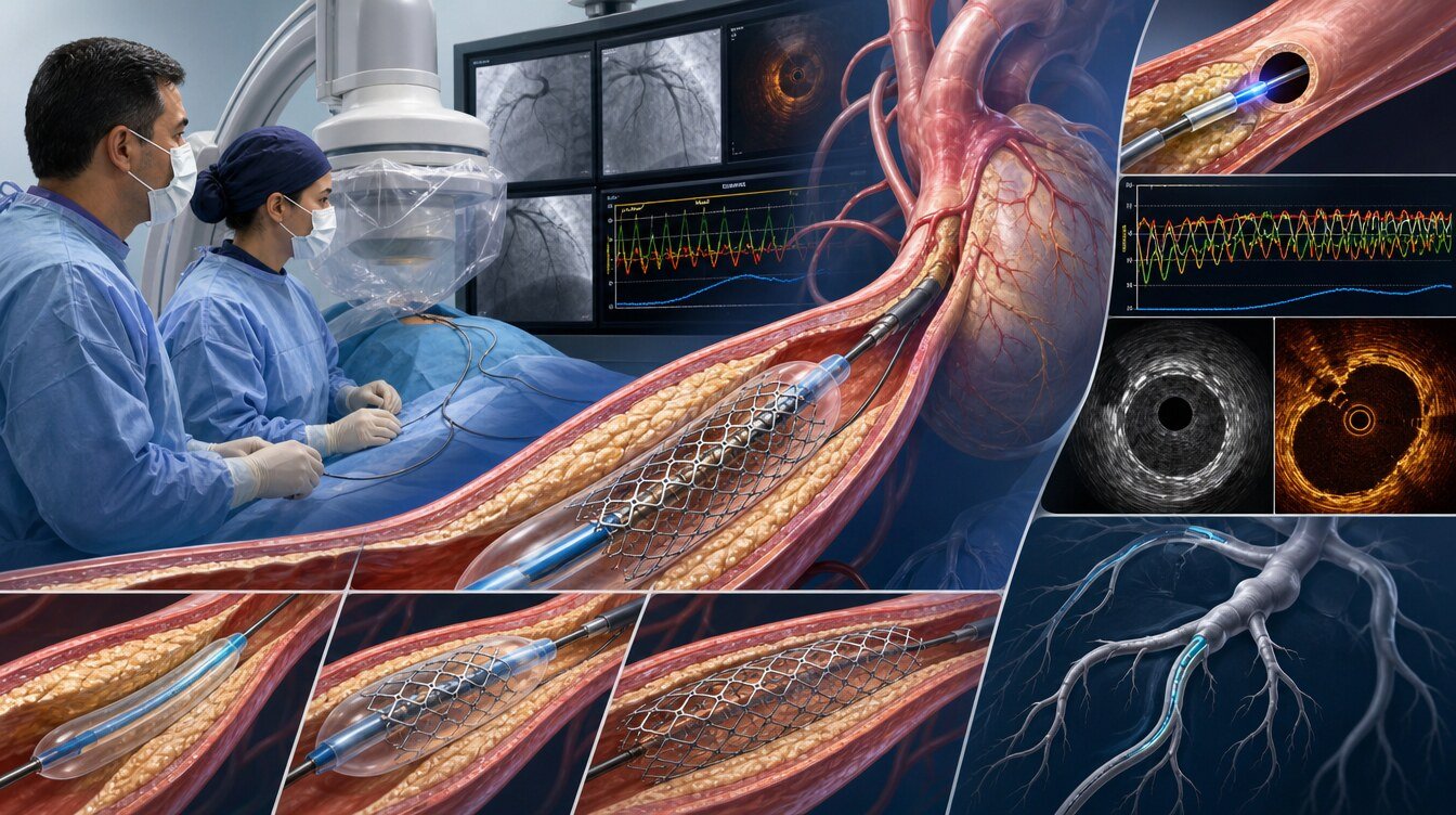
- FFR uses a pressure wire to assess how much a lesion affects flow.
- iFR measures pressure during a defined part of the cardiac cycle and usually needs no vasodilator.
- IVUS uses ultrasound inside the vessel to image the wall, plaque and stent expansion.
- OCT uses high-resolution light-based imaging to assess stent apposition and vessel detail.
FFR/iFR mainly assess functional significance; IVUS/OCT assess anatomical features. In complex bifurcations, OCT-guided PCI reduced two-year major cardiac events in OCTOBER. In ILUMIEN IV, OCT produced a larger stent area and less stent thrombosis, but not a significant difference in the two-year composite target-vessel endpoint.[11,12]
How is a stent procedure performed?
The procedure is performed in a catheter laboratory. The usual steps are:
- Preparation: Blood pressure, rhythm and oxygen are monitored; IV access and blood tests are checked.
- Arterial access: Local anaesthetic is applied around the radial artery at the wrist or femoral artery in the groin.
- Angiography: A thin catheter images the coronary arteries and defines the lesion.
- Guidewire: A very fine wire is advanced beyond the narrowing.
- Lesion preparation: Balloon, cutting/scoring balloon, atherectomy or intravascular lithotripsy may be used; severe calcium may require calcified lesion treatment.
- Stent expansion: The balloon-mounted stent is expanded; an additional balloon may optimize it.
- Final check: Flow and placement are checked by angiography and, in selected cases, IVUS/OCT.
- Access closure: Wrist compression or groin compression/closure is applied.
Most patients remain awake. Duration varies with lesion number and complexity. A simple PCI is not equivalent to bifurcation/LMCA intervention, chronic total occlusion PCI or treatment of severe calcification.
Is stenting performed through the wrist or groin?
Radial access is preferred in suitable patients, especially in acute coronary syndromes, because it reduces bleeding and access-site complications.[1,2] Femoral access may be more suitable when large equipment is needed, wrist vessels are unsuitable, complex procedures require support or individual anatomy dictates it. Access is selected for the procedure and patient.
Stent or bypass surgery?
PCI and bypass are not simple substitutes. Left-main involvement; number, location and complexity of lesions; diabetes; heart function; prior procedures; surgical risk, age and frailty; kidney, lung and vascular disease; and patient preferences matter.
The 2024 ESC guideline generally identifies bypass as preferred for low-surgical-risk left-main disease, and bypass is favoured in some groups with multivessel disease and diabetes. PCI may be considered in patients unsuitable for surgery or at high risk and in selected low-to-intermediate complexity anatomy.[3,4] Complex decisions benefit from a Heart Team and shared decision-making.[3–5,23]
Which medicines are used after stenting?
Why is antiplatelet therapy important?
Dual antiplatelet therapy (DAPT), commonly aspirin plus a P2Y12 inhibitor, is generally prescribed to prevent platelets forming a clot on the stent. Do not stop these medicines without medical advice. Early interruption can increase stent thrombosis and heart-attack risk. If bleeding, surgery or dental treatment arises, the cardiology and procedural teams should decide together.
How long does DAPT continue?
- Acute coronary syndrome: The 2025 ACC/AHA default is at least 12 months without high bleeding risk; shorter or different strategies may be considered when bleeding risk is high.[1]
- Chronic coronary syndrome: The 2024 ESC guideline generally individualizes 1–6 months according to bleeding and thrombotic risk.[3]
- Long-term anticoagulation: The period of triple therapy is minimized to reduce bleeding and is individualized.[1,3]
MASTER DAPT supported abbreviated therapy after one month in defined high-bleeding-risk patients; TWILIGHT found less bleeding with ticagrelor alone after three event-free months in selected high-risk patients.[13,14] These findings do not mean every patient can stop medication early.
Other treatment
Stenting is only one part of long-term atherosclerosis treatment. Lipid lowering, blood-pressure and diabetes control, smoking cessation, anti-anginal therapy, nutrition, physical activity and cardiac rehabilitation may be recommended.[1–4,17]
What are the risks of stenting?
PCI is common but not risk-free. Risk varies with urgency, age, kidney function, bleeding risk, anatomy and complexity.
- Bleeding, bruising or vessel injury at the wrist or groin,
- Contrast reaction or kidney injury,
- Vessel-wall tear or abrupt closure, rhythm disturbance, periprocedural heart attack or stroke,
- Emergency bypass, stent thrombosis, restenosis and, very rarely, death.
A simple planned single-vessel radial PCI does not carry the same risk as complex multivessel PCI during a heart attack.
What is stent thrombosis?
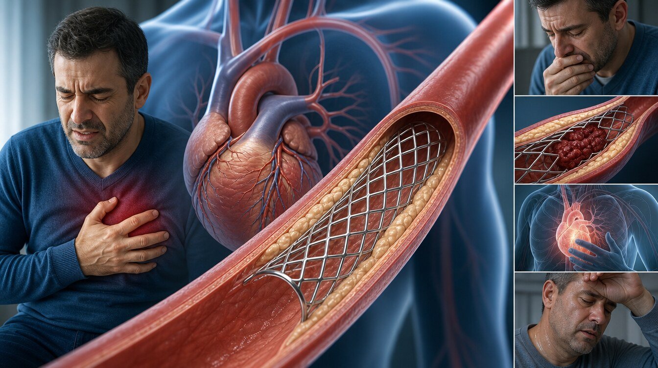
It is clot formation inside the stent. It commonly causes sudden chest pain and a heart attack and requires emergency treatment. Early antiplatelet interruption, inadequate expansion, complex lesions and individual thrombotic tendency can affect risk.
What is in-stent restenosis?
It is recurrent narrowing due to excessive tissue growth or disease progression. It is usually less abrupt than thrombosis and may cause exertional pain or breathlessness. Depending on intravascular imaging, treatment may include a drug-coated balloon, another stent or a different intervention.
How long is the hospital stay after stenting?
Same-day discharge may be possible after uncomplicated planned PCI in suitable patients, but it is not universal. Successful completion without bleeding or early complications; stable vital signs, access site and comorbidities; and adult support at home, emergency access, medicine supply and follow-up are assessed.[15,16] Heart attack, complex PCI, ongoing pain, rhythm problems, kidney risk or access complications may require longer observation.
Returning to daily life after stenting
The discharge document and treating team's instructions take priority. Timing varies with access, heart attack, wound healing and occupation.
Walking and exercise
Light walking can often begin early. Heavy lifting and strenuous exercise wait until the access site heals; some sources advise avoiding heavy lifting for about a week after uncomplicated elective PCI. Return after a heart attack is more gradual.[17,18] Cardiac rehabilitation combines exercise, risk-factor education, medication adherence, nutrition and psychosocial support.[1,4,18]
Work and driving
Some people return to desk work in about a week after uncomplicated elective PCI; physical work may take longer.[17] After a heart attack, heart function, symptoms and licence class matter. These estimates are not universal legal rules; obtain written personal advice.
Air travel
Some centres advise delaying flying for 1–2 weeks after uncomplicated elective PCI; heart attack, heart failure or complications may extend this.[18] Check with the cardiology team, airline and insurer.
Can I have an MRI?
Most modern stents are MR Conditional. Safety depends on model and scanner conditions. Keep the stent card and give the MRI unit the implant date and product details.[24,25]
Dental treatment or surgery
Timing depends on why the stent was placed and whether antiplatelets must be interrupted. The 2024 AHA/ACC guideline recommends ideally delaying elective surgery requiring interruption for at least 12 months after DES for ACS and at least six months after DES for chronic disease; time-sensitive surgery may be considered after three months in selected patients by multidisciplinary decision.[5] A dentist or surgeon should not independently stop antiplatelet therapy.
Which symptoms need emergency help?
- Chest pain that persists at rest, worsens or resembles prior cardiac pain,
- Chest pain with breathlessness, cold sweat, nausea or fainting,
- Access-site bleeding despite pressure or rapidly enlarging swelling,
- Coldness, pallor, blue colour, numbness or weakness in the accessed limb,
- Sudden speech difficulty, face/arm weakness, severe palpitations or severe breathlessness.
Call 112 or local emergency services. Minor bruising and tenderness can be common; contact the procedural centre for increasing pain/swelling, redness, discharge or fever.[17]
Why does follow-up continue after stenting?
A stent opens only the treated segment; atherosclerosis may progress elsewhere. Follow-up reviews symptoms and exercise capacity, blood pressure, LDL and other laboratory values, diabetes, adherence and bleeding, smoking, nutrition, activity and rehabilitation. Hyperlipidemia treatment is central to secondary prevention.
Routine repeat angiography in an asymptomatic patient is not required for everyone. New or progressive symptoms, a high-risk test or a specific clinical reason may prompt further assessment.[3,4]
Prof. Dr. Hakan Uçar's scientific work in this field
Work directly related to coronary stenting and PCI includes a multicentre observational comparison of crossover versus focal ostial LAD stenting; a 64-patient experience with the jailed semi-inflated balloon technique in bifurcations; a 64-patient technical/observational study of the modified flower petal technique; and a case report of stent thrombosis in rare coronary anatomy.[19–22]
These publications demonstrate academic work but do not replace guidelines or large randomized trials. Observational and technical studies and case reports should not be presented as evidence of causal superiority.
Frequently asked questions
No. It is a percutaneous procedure performed with catheters, usually through a wrist or groin artery. The chest is not opened.
Current metal drug-eluting stents remain permanently in the artery and gradually become covered by vessel tissue.
No. A drug-eluting stent leaves a scaffold; a drug-coated balloon transfers medication and is removed. Suitability depends on the lesion.
Brief, different discomfort may occur, but persistent, worsening or familiar cardiac pain needs emergency assessment.
Clot or gradual restenosis can occur. Modern stents and appropriate medication reduce but do not eliminate risk.
Contact the cardiology team or prescriber that day. Do not independently double a dose or stop treatment.
Timing depends on procedure indication, heart function, access site and licence class. Obtain advice specific to local rules and your condition.
Most dental procedures can be planned, but antiplatelet management is crucial. Do not stop medication without joint dental and cardiology advice.
Assessment and decision
Coronary stenting can be lifesaving in the right acute setting and can reduce symptoms and some ischaemic events in selected stable patients. The decision is not based on a percentage narrowing alone; symptoms, flow assessment, anatomy, medical therapy, bleeding risk and bypass options are considered together.
Literature
References
Rao SV, O'Donoghue ML, Ruel M, et al. 2025 ACC/AHA/ACEP/NAEMSP/SCAI Guideline for Acute Coronary Syndromes. Circulation. 2025. DOI: 10.1161/CIR.0000000000001309
Byrne RA, Rossello X, Coughlan JJ, et al. 2023 ESC Guidelines for acute coronary syndromes. European Heart Journal. 2023;44:3720–3826. DOI: 10.1093/eurheartj/ehad191
Vrints C, Andreotti F, Koskinas KC, et al. 2024 ESC Guidelines for chronic coronary syndromes. European Heart Journal. 2024;45:3415–3537. DOI: 10.1093/eurheartj/ehae177
Virani SS, Newby LK, Arnold SV, et al. 2023 AHA/ACC Guideline for Chronic Coronary Disease. Circulation. 2023;148:e9–e119. DOI: 10.1161/CIR.0000000000001168
Thompson A, Fleischmann KE, Smilowitz NR, et al. 2024 AHA/ACC Perioperative Cardiovascular Management Guideline. Circulation. 2024;150:e351–e442. DOI: 10.1161/CIR.0000000000001285
Boden WE, O'Rourke RA, Teo KK, et al. Optimal medical therapy with or without PCI for stable coronary disease. New England Journal of Medicine. 2007;356:1503–1516. DOI: 10.1056/NEJMoa070829
Maron DJ, Hochman JS, Reynolds HR, et al. Initial invasive or conservative strategy for stable coronary disease. New England Journal of Medicine. 2020;382:1395–1407. DOI: 10.1056/NEJMoa1915922
Xaplanteris P, Fournier S, Pijls NHJ, et al. Five-year outcomes with FFR-guided PCI. New England Journal of Medicine. 2018;379:250–259. DOI: 10.1056/NEJMoa1803538
Al-Lamee R, Thompson D, Dehbi HM, et al. PCI in stable angina (ORBITA). Lancet. 2018;391:31–40. DOI: 10.1016/S0140-6736(17)32714-9
Rajkumar CA, Foley MJ, Ahmed-Jushuf F, et al. Placebo-controlled PCI for stable angina (ORBITA-2). New England Journal of Medicine. 2023;389:2319–2330. DOI: 10.1056/NEJMoa2310610
Ali ZA, Landmesser U, Maehara A, et al. OCT-guided versus angiography-guided PCI (ILUMIEN IV). New England Journal of Medicine. 2023;389:1466–1476. DOI: 10.1056/NEJMoa2305861
Holm NR, Andreasen LN, Neghabat O, et al. OCT or angiography guidance in bifurcation PCI (OCTOBER). New England Journal of Medicine. 2023;389:1477–1487. DOI: 10.1056/NEJMoa2307770
Valgimigli M, Frigoli E, Heg D, et al. DAPT after PCI in high bleeding risk (MASTER DAPT). New England Journal of Medicine. 2021;385:1643–1655. DOI: 10.1056/NEJMoa2108749
Mehran R, Baber U, Sharma SK, et al. Ticagrelor with or without aspirin after PCI (TWILIGHT). New England Journal of Medicine. 2019;381:2032–2042. DOI: 10.1056/NEJMoa1908419
Rao SV, Vidovich MI, Gilchrist IC, et al. ACC Consensus on Same-Day Discharge After PCI. Journal of the American College of Cardiology. 2021;77:811–825. DOI: 10.1016/j.jacc.2020.11.013
Seto AH, Shroff A, Abu-Fadel M, et al. Length of stay following PCI: SCAI update. Catheterization and Cardiovascular Interventions. 2018;92:717–731. DOI: 10.1002/ccd.27637
NHS. Coronary angioplasty and stent insertion — Recovery. Accessed 25 August 2026.
American Heart Association. What is Cardiac Rehabilitation? Accessed 25 August 2026.
Soylu K, Yildirim U, Nasifov M, Uçar H, et al. Crossover or focal ostial LAD stenting. Anatolian Journal of Cardiology. 2022;26:827–831. DOI: 10.5152/AnatolJCardiol.2022.1122
Ermiş E, Uçar H, Demirelli S, et al. Jailed semi-inflated balloon technique in bifurcation lesions. Türk Kardiyoloji Derneği Arşivi. 2018;46:340–348. DOI: 10.5543/tkda.2018.47347
Çaylı M, Elbasan Z, Gür M, Şeker T, Uçar H, et al. Modified flower petal technique. EuroIntervention. 2015;11:772–779. DOI: 10.4244/EIJV11I7A154
Ermiş E, Kahraman S, Uçar H, et al. Stent thrombosis in anomalous coronary anatomy: case report. Intractable & Rare Diseases Research. 2018;7:54–57. DOI: 10.5582/irdr.2018.01001
Lawton JS, Tamis-Holland JE, Bangalore S, et al. 2021 ACC/AHA/SCAI Coronary Revascularization Guideline. Circulation. 2022;145:e18–e114. DOI: 10.1161/CIR.0000000000001038
U.S. Food and Drug Administration. Engineering Tests and Recommended Labeling for Intravascular Stents. Accessed 25 August 2026.
U.S. Food and Drug Administration. Coronary stent patient information: MRI conditions on implant card. Accessed 25 August 2026.
Continue