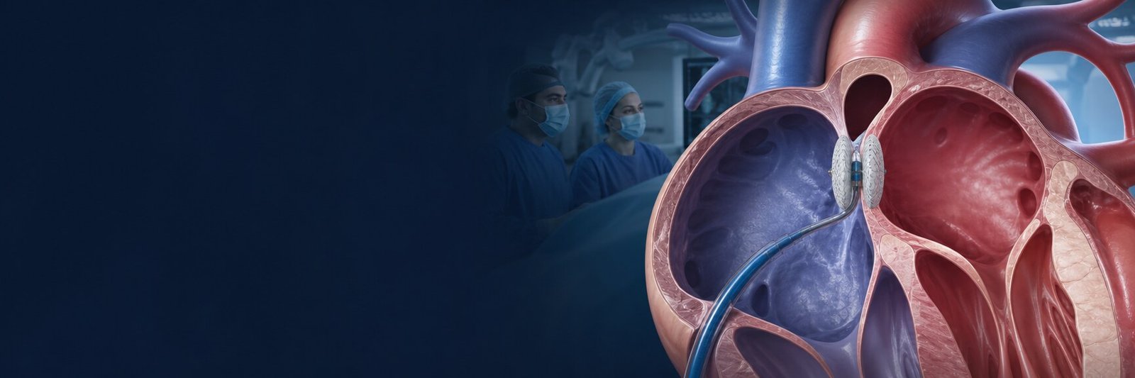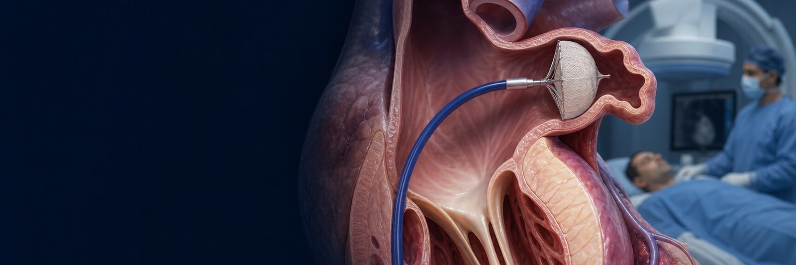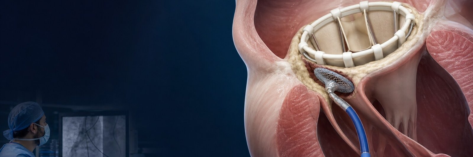
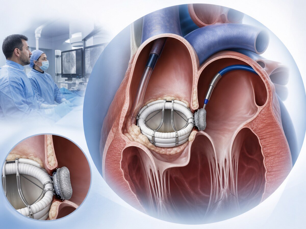
PVL closure is reduction of a leak around a prosthetic heart valve with a catheter-placed closure device in suitable people. The name means paravalvular leak closure. PVL is flow around the suture line or landing zone, not through the prosthesis itself. It can appear after surgical mitral or aortic valve replacement and after some transcatheter valve procedures. PVL can be found in about 5–17% of surgical prostheses; only about 2–5% cause clinical problems such as heart failure or haemolytic anaemia.[4]
A catheter-based method can be done without opening the chest and often without a further open-heart operation, but it is not suitable for every person or every leak. Imaging, symptoms, the degree of anaemia, the state of the prosthesis and surgical risk are weighed together.[1,2]
How does PVL closure differ from other procedures?
PVL closure is one of the transcatheter closures in structural heart interventions, but the clinical question is different.
- PFO closure and ASD closure: they close a passage or opening in the wall between the atria. They do not treat leak around a prosthetic valve.
- LAA closure: occludes the left atrial appendage for stroke prevention; it is not PVL closure.
- TAVI and heart-valve interventions: they implant a new valve or treat valve disease. Paravalvular flow can appear after a transcatheter valve; that does not make those procedures the same as PVL closure.[3]
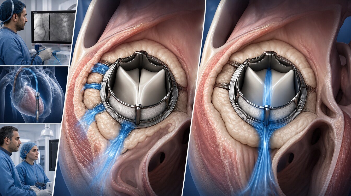
Why does a leak form around a prosthetic valve?
After surgical mitral and aortic valves
In surgical valve replacement the prosthesis is sewn to heart tissue. Over time the suture line can loosen, the tissue can weaken, calcium can form, healing can be incomplete, or the prosthesis and the tissue can be mismatched. Blood then leaks between chambers around the valve; that is PVL.[3]
In mitral PVL the leak sits around a mitral prosthesis between the left atrium and the left ventricle. In aortic PVL it sits around an aortic prosthesis between the left ventricle and the aorta. Mitral leaks are often more complex for catheter access and device seating; calcified leak channels and a mitral site have been reported among risk factors linked to haemolysis.[7]
After transcatheter valves
Leak between a transcatheter valve and the native annulus can also occur. Some degree of paravalvular prosthetic leak has been found in about 5–10% of surgical prostheses and about 40–70% of transcatheter valves. Those figures describe any paravalvular flow seen on imaging, not clinically important leak; many people have no symptoms and no important cardiac load.[3]
Active or previous prosthetic-valve infection can damage tissue around the valve and loosen the prosthesis. New or rapidly increasing leak therefore needs an infection work-up.[1]
What symptoms can PVL cause?
The importance of a leak is not set by image size alone. Site, shape, pressure and volume load, pumping strength and the person’s overall health also matter. Clinically important PVL can cause:
- breathlessness on effort, or new breathlessness
- difficulty breathing when lying flat
- waking at night with breathlessness
- swelling of the legs or ankles
- easy fatigue and a reduced exercise capacity
- palpitations
- chest pressure or discomfort
- abdominal bloating or a rapid weight gain
- dizziness or fainting
- pallor, fatigue and palpitations on effort from anaemia
When heart failure develops, the leak can volume-load the chambers. Over time the heart can enlarge, fluid can collect in the lungs, and breathlessness can require hospital admission.[4,9]
Breathlessness is not caused by PVL alone. Anaemia, arrhythmia, lung disease, kidney problems or another prosthetic-valve problem can look similar. Symptoms are therefore read with the examination and tests.
Haemolysis, haemolytic anaemia and transfusion
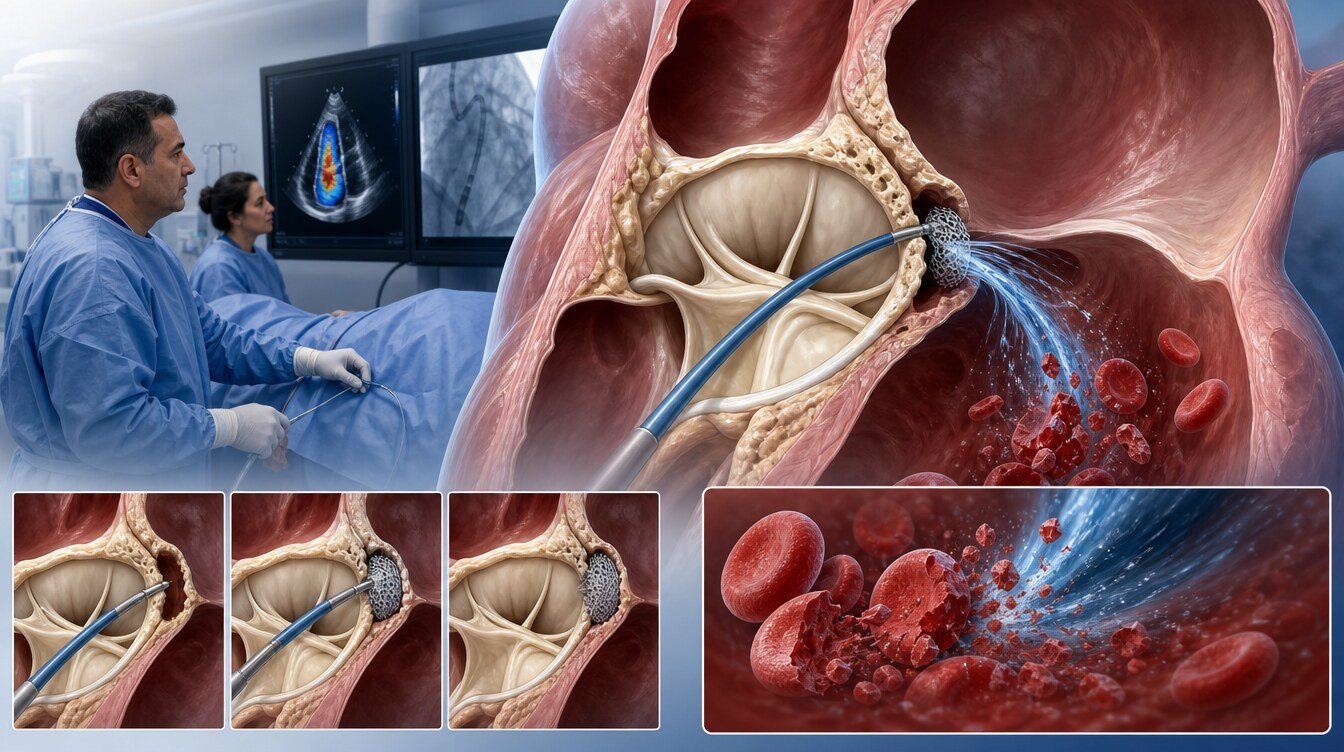
High-velocity, turbulent flow through a PVL can damage red blood cells. That is haemolysis. When it is marked, haemolytic anaemia can follow. Haemoglobin falls; lactate dehydrogenase may rise, haptoglobin may fall, and blood or urine can show red-cell breakdown.[8]
Symptoms can include fatigue, breathlessness, palpitations, dizziness and pallor. Some people notice jaundice or darker urine. Iron or other support, or blood transfusion, may be needed depending on the anaemia and the heart and lungs. In one study, transfusion was needed in 34% of people before PVL closure and in 21% afterwards; haemolysis markers did not improve in everyone. Those percentages are not a personal success rate.[8]
Closing the leak can reduce haemolysis. In a prospective registry, closing at least 90% of the leak’s cross-sectional area was linked to less haemolysis, and that effect lasted at 6 months. Incomplete closure can leave haemolysis unchanged or increase it in some people.[7] What matters is not only that a device was placed, but how much leak remains and how red-cell breakdown behaves in follow-up.
How is PVL separated from other prosthetic-valve problems?
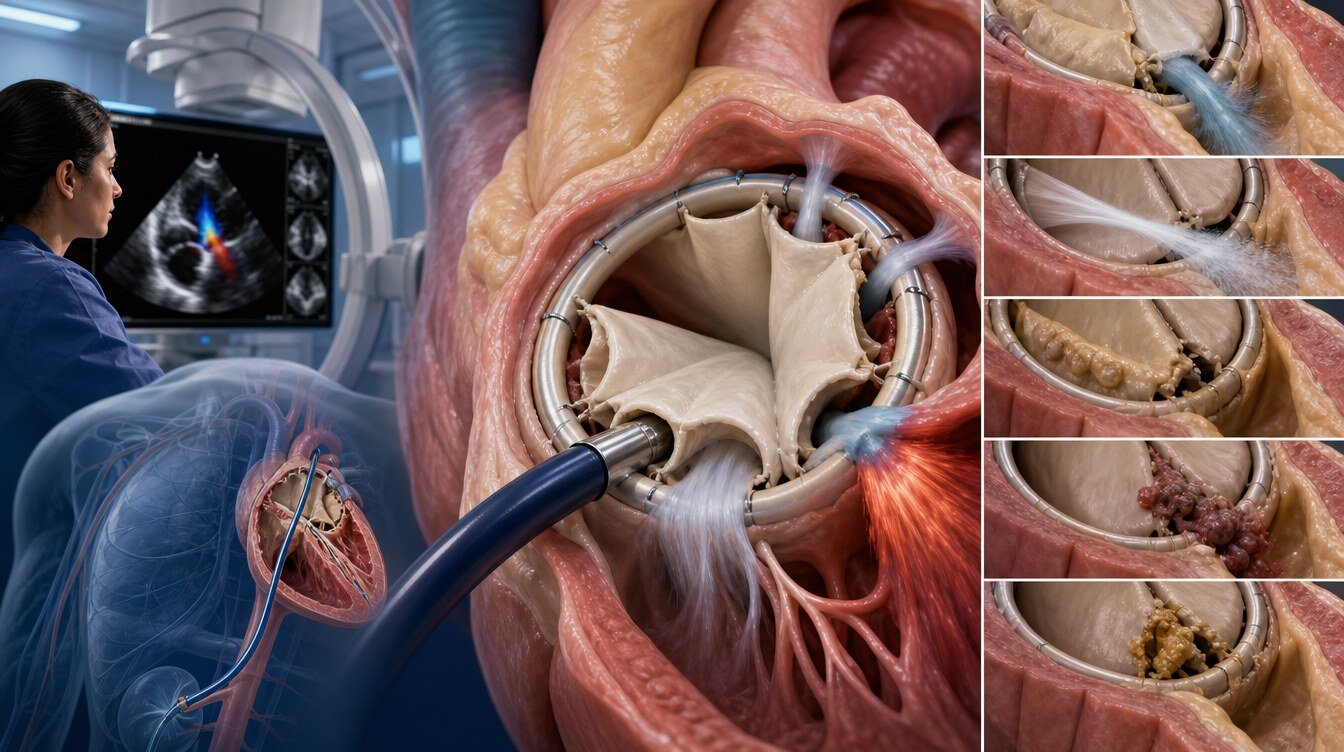
Normal “washing jets”
Especially with mechanical valves, very small, brief jets can appear around the moving parts. These can be normal “washing jets” built into the design and do not mean PVL. Site, direction, severity and prosthetic function are reviewed. Colour Doppler flow alone does not mean closure is needed.[1]
Transvalvular prosthetic leak
Transvalvular leak is blood returning through or between the moving parts of the prosthesis. PVL comes from the gap between the outer edge of the prosthesis and the heart tissue. Treatment and the procedure plan can differ. Echocardiography is used to see where the jet starts.[11]
Structural valve degeneration
Biological prostheses can thicken, calcify, wear or tear over time. That is structural valve degeneration. The problem then sits in the prosthesis itself; in PVL the main problem is the suture line or annulus. Both can be present in the same person.
Prosthetic-valve thrombosis
Clot can form on or around the prosthesis and restrict leaflet or disc motion. This can relate to under-anticoagulation, especially with mechanical valves, or to other risk factors. Suspected thrombosis is not managed as PVL closure; anticoagulation, further imaging or, in selected cases, another intervention may be needed.[2]
Active infective endocarditis
Active infective endocarditis is a serious infection of a heart valve or the tissue around a prosthesis. Fever, chills, night sweats, unexplained fatigue or a new murmur can appear. Closing the leak with a device during active infection does not treat the infection and may be inappropriate. Blood cultures, echocardiography and review by infectious diseases and cardiac surgery are needed.[1]
Finding a leak does not automatically mean closure
Seeing PVL on imaging is not, by itself, a reason to intervene. Mild leaks that cause no symptoms, no important chamber load and no haemolysis can be followed with clinical and echocardiographic review. The decision weighs severity, symptoms, heart failure, haemolysis, prosthetic stability and the chance of infection.[1,2]
Who may be considered for PVL closure?
Transcatheter PVL closure may be considered when one or more of the following is present:
- Breathlessness, oedema or recurrent heart-failure findings thought to be due to the leak
- PVL confirmed as important on imaging
- Haemolytic anaemia related to the leak
- Ongoing or recurrent need for transfusion
- Redo surgery that carries high or prohibitive risk
- Anatomy that can be reached with a catheter and can accept a device
The catheter option is often considered when repeat sternotomy or further valve surgery is high or prohibitive risk. The decision is not made from age or a single risk score alone.[1,4]
When is it not a routine catheter option?
Transcatheter PVL closure is not a routine approach when:
- the leak is mild and there are no symptoms
- there is active infective endocarditis
- the anatomy is too irregular, too large or too hard to reach for safe device seating
- the prosthesis is loose or mechanically unstable
- the device is likely to restrict leaflet or disc motion
- the leak is in fact caused by thrombosis, structural degeneration or transvalvular regurgitation
Surgery to repair or replace the prosthesis may then be needed. If infection is active, treating the infection comes first; the procedure decision is made with infection control, heart failure and prosthetic stability.[1]
Surgical and transcatheter closure: which option, and when?
Redo surgery
Surgery can repair the leak directly or replace the prosthesis when needed. It may be required if the prosthesis is loose, the valve structure is damaged, endocarditis is active, the leak is very large or multiple, or a catheter device is likely to interfere with valve function. Technical success can be higher, but redo surgery carries risks of bleeding, infection, kidney injury, stroke and early death.[5,6]
Transcatheter closure
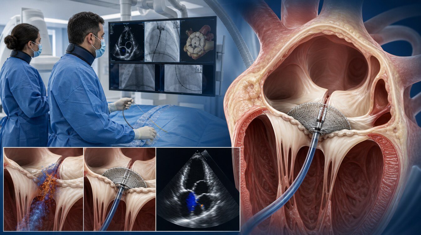
In a transcatheter procedure, one or more closure devices are placed in the PVL through catheters. It may be considered when redo surgery is high risk, the leak causes symptoms or haemolysis, and the anatomy is suitable. Experienced centres have reported procedural success around 90%; that rate varies with case mix, leak anatomy and technique and is not a personal success promise.[3]
Most comparative studies are retrospective and observational. Technical success has been higher with surgery, while early morbidity and death can be higher; mid-term death and some clinical-effect measures have been similar in some series.[4,5] No randomised trial has compared surgery with transcatheter PVL closure directly.[4] There is therefore no single “better for everyone” method.
How is the decision made? Heart Team and shared decision-making
PVL treatment usually needs cardiology, interventional cardiology, cardiac surgery, imaging, anaesthesia and, when needed, infectious diseases. That group is the Heart Team.[3]
The review asks:
- Is the jet truly PVL?
- What is its grade and site?
- Can the symptoms be linked to the leak?
- Is there heart failure or haemolysis?
- Has active infection been excluded?
- Is the prosthesis stable and working?
- What is the estimated risk of redo surgery?
- Is a catheter path and device seat technically possible?
- What are the risks if no treatment is given?
- What are the person’s preferences and life goals?
The likely benefits, limits, alternatives and the path if no procedure is done should be explained. The final decision joins the medical review with the person’s informed preference.[1,2]
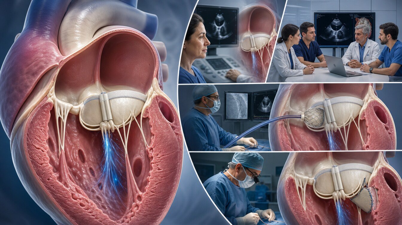
Diagnosis and imaging
Transthoracic echocardiography
TTE is ultrasound from the chest wall. It helps review pumping strength, chamber size, prosthetic motion and the pressure–volume load from the leak. Acoustic shadowing from the prosthesis can hide small or eccentric jets.[12]
Two- and three-dimensional transoesophageal echocardiography
TEE uses a probe in the oesophagus. Being closer to the heart, it can show the site, shape and width of the PVL and prosthetic motion in more detail. Three-dimensional colour Doppler TEE helps measure the leak area and the device’s relation to neighbouring tissue; it is also used during the procedure to seat the device and check residual leak.[10,11]
Fluoroscopy and cardiac computed tomography
Fluoroscopy can follow mechanical discs or leaflets and the catheters. Cardiac CT can show the sewing ring, the leak channel, calcium and neighbouring anatomy in three dimensions. TEE and CT together can make pre-procedure planning more detailed.[12,13]
Electrocardiography, blood counts, kidney function, haemolysis tests, blood cultures and coronary assessment are added when needed. Not every test is needed in every person; tests follow the clinical picture and the planned procedure.
How is transcatheter PVL closure performed?
The procedure is usually done in a catheter laboratory that combines angiography with advanced echocardiography. The anaesthetic plan depends on duration, the person’s condition and the leak site. General anaesthesia or other sedation options may be used.
Access routes
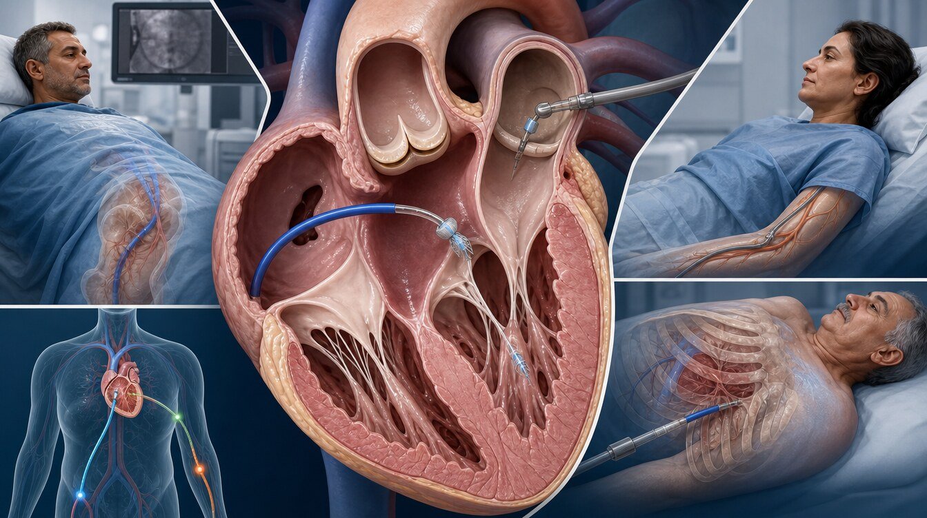
Access is individualised to the leak site and the prosthetic anatomy. Routes can include:
- Transseptal access: crossing the wall between the atria, especially for mitral PVL
- Retrograde arterial access: reaching the heart from the aorta
- Transapical access: a dedicated approach through the left-ventricular apex
- Other specialised or hybrid routes: alternatives set by the person’s anatomy
The same route is not used in every person. The plan is set from CT and TEE, previous operations and the team’s review.[13,14]
How is the closure device chosen?
Device choice follows leak length, width, shape, channel direction, nearby calcium, distance to the moving parts of the prosthesis, and whether more than one leak is present. More than one device may be needed. The aim is to reduce the leak without restricting prosthetic opening and closing. Undersizing on three-dimensional imaging has been linked to failed closure, so measurement and device size are planned carefully.[10]
This is not open-heart surgery. Avoiding a chest incision does not make the procedure risk-free or suitable for everyone. Duration, fasting time or discharge day should not be copied from this page.
Risks and complications
PVL closure is not risk-free even when planned by experienced teams. Reported or clinically relevant complications include:
- incomplete closure or residual leak
- the device restricting leaflet or disc motion
- device migration or embolisation
- vessel injury, bleeding or haematoma
- perforation of the heart wall or neighbouring tissue
- bleeding into the pericardium and tamponade
- stroke or systemic embolism
- contrast allergy
- a decline in kidney function
- arrhythmia or conduction problems
- ongoing or increased haemolysis
- infection
- the need for emergency surgery
- readmission or death
Risk varies with leak site, procedure time, access route, kidney function, previous operations, prosthetic design and overall health. Series of percutaneous procedures have reported technical difficulty, long procedure times and complications.[3] These risks should be explained in person before the procedure.
Recovery after the procedure
Heart rhythm, bleeding, the access site, prosthetic motion and residual leak are watched, including with echocardiography. Length of stay depends on the procedure, the access route, complications and other illness. Observational comparisons have found a shorter stay after transcatheter treatment than after surgery; that does not apply in the same way to every person.[4,5]
After going home, growing swelling, bleeding, redness or pain at the access site, fever, new breathlessness, chest pain, fainting, weakness or speech difficulty need attention. Return to daily activity depends on the access site and recovery. Medicines should not be stopped or changed without medical advice.
Antithrombotic treatment and follow-up
There is no single antithrombotic plan after PVL closure. Treatment is individualised to a mechanical or biological prosthesis, atrial fibrillation, previous clot or embolism, bleeding risk, the closure device and other medicines.[2,15]
Follow-up reviews symptoms, heart-failure findings, blood counts, haemolysis markers and prosthetic function. TTE is the usual first test; TEE, CT or other imaging is added when needed. Intervals follow personal risk and the procedural result.
When is urgent review needed?
Urgent or same-day medical review is needed for:
- new or rapidly worsening breathlessness
- inability to lie flat, a choking feeling at night, or pink frothy sputum
- fainting or new marked dizziness
- severe chest pain
- one-sided weakness, facial droop or speech difficulty
- high fever, chills or unexplained ongoing fatigue
- clearly darker urine or jaundice
- rapidly increasing pallor, palpitations or haemolysis findings
- bleeding that will not stop, or rapidly growing swelling at the access site
Fever or chills should prompt a review for prosthetic-valve infection. Blood cultures may be needed before antibiotics; that decision is made by a clinician. Infection concern in a person with a prosthetic valve should not be delayed.[1] In Türkiye, call 112 for urgent symptoms.
Residual leak and ongoing haemolysis
The leak may not disappear completely. A small residual jet can be clinically unimportant; in some people leftover flow can keep heart-failure symptoms or haemolysis going. Success is therefore not judged only as “a device was placed”; the effect of leftover leak on prosthetic function, cardiac load and blood values is also followed.[9]
If haemoglobin stays low, LDH stays high or transfusion is still needed, echocardiography and haemolysis tests can be repeated. A second catheter procedure, a different device, surgery or supportive care may then be considered. Incomplete closure has been reported to increase haemolysis in some people.[7]
What happens if PVL is not closed?
Mild, clinically unimportant PVL can be followed for years without a major problem. An important untreated leak can increase volume load, enlarge the chambers, worsen heart failure, lead to repeat admissions or cause haemolytic anaemia.[3,9]
If haemolysis continues, anaemia can deepen and some people need transfusion. If the leak is linked to active infection, the infection can progress and the prosthesis can loosen. Immediate closure is still not right for every PVL; follow-up, medicines, infection treatment, surgery or a catheter option are chosen for the person.[1]
Frequently asked questions
No. Transcatheter PVL closure uses catheters and closure devices advanced through blood vessels. Avoiding a chest incision does not make the procedure risk-free or suitable for every person.[3]
No. Mild leaks that cause no symptoms and no haemolysis can be followed. The decision depends on imaging, heart failure, haemolysis and the state of the prosthesis.[1]
No. PVL can occur after surgical and transcatheter valves. Paravalvular flow is reported more often after transcatheter valves, but not every jet is clinically important.[3]
High-velocity, turbulent flow through the leak can damage red blood cells. That mechanical injury can cause haemolysis, a lower haemoglobin and haemolytic anaemia.[8]
Not reliably. Closing most of the leak can reduce haemolysis, but residual flow, more than one leak, device position or other causes can leave anaemia unchanged.[7]
Yes. If the device sits close to the leaflets or mechanical discs, it can restrict motion. Device choice and placement are therefore checked with detailed imaging.[10]
No. Anticoagulant or antiplatelet treatment is set by the type of prosthesis, heart rhythm, clot and bleeding risk, and the device used. Medicines should not be stopped without medical advice.[2]
In a person with a prosthetic valve, fever, chills or unexplained fatigue should be assessed for infection. New or ongoing fever needs prompt medical review; blood cultures and echocardiography may be required.[1]
Assessment and decision
The PVL closure decision does not rest on the sentence “there is a leak on imaging, so close it.” Whether the jet is truly paravalvular, symptoms, haemolysis, infection, prosthetic stability, surgical risk, catheter anatomy and the person’s preferences are read together. Much of the evidence is observational; no randomised trial has compared surgical and transcatheter PVL closure directly.[1,2,4]
Literature
References
Praz F, et al. 2025 ESC/EACTS Guidelines for the management of valvular heart disease. European Heart Journal. 2025. doi:DOI: 10.1093/eurheartj/ehaf194.
Otto CM, et al. 2020 ACC/AHA Guideline for the Management of Patients With Valvular Heart Disease: Executive Summary. Journal of the American College of Cardiology. 2021. doi:DOI: 10.1016/j.jacc.2020.11.035.
Sorajja P, et al. Percutaneous repair of paravalvular prosthetic regurgitation: patient selection, techniques and outcomes. Heart. 2015. doi:DOI: 10.1136/heartjnl-2014-306270.
Molina Ramos AI, et al. Paravalvular leaks: short and long-term outcomes of surgical versus percutaneous closure. European Heart Journal. 2021. doi:DOI: 10.1093/eurheartj/ehab724.2238.
Busu T, et al. Meta-analysis comparing transcatheter and surgical treatments of paravalvular leaks. The American Journal of Cardiology. 2018. doi:DOI: 10.1016/j.amjcard.2018.03.360.
Millán X, et al. Surgery Versus Transcatheter Interventions for Significant Paravalvular Prosthetic Leaks. JACC: Cardiovascular Interventions. 2017. doi:DOI: 10.1016/j.jcin.2017.08.013.
Smołka G, et al. Transcatheter paravalvular leak closure and hemolysis – a prospective registry. Archives of Medical Science. 2016. doi:DOI: 10.5114/aoms.2016.60435.
Panaich SS, et al. Effect of percutaneous paravalvular leak closure on hemolysis. Catheterization and Cardiovascular Interventions. 2018. doi:DOI: 10.1002/ccd.27917.
Ruiz CE, et al. Clinical outcomes in patients undergoing percutaneous closure of periprosthetic paravalvular leaks. Journal of the American College of Cardiology. 2011. doi:DOI: 10.1016/j.jacc.2011.03.074.
Franco E, et al. Three-Dimensional Color Doppler Transesophageal Echocardiography for Mitral Paravalvular Leak Quantification and Evaluation of Percutaneous Closure Success. Journal of the American Society of Echocardiography. 2014. doi:DOI: 10.1016/j.echo.2014.08.019.
Lázaro C, et al. Cardiac imaging in prosthetic paravalvular leaks. Cardiovascular Diagnosis and Therapy. 2014. doi:DOI: 10.3978/j.issn.2223-3652.2014.07.01.
O’Neill AC, et al. Practical tips and tricks for assessing prosthetic valves and detecting paravalvular regurgitation using cardiac CT. Journal of Cardiovascular Computed Tomography. 2014. doi:DOI: 10.1016/j.jcct.2014.07.001.
Garcia-Sayan E, et al. Multimodality cardiac imaging for procedural planning and guidance of transcatheter mitral valve replacement and mitral paravalvular leak closure. Frontiers in Cardiovascular Medicine. 2021. doi:DOI: 10.3389/fcvm.2021.582925.
Desai A, et al. Update in paravalvular leak closure. Current Cardiology Reports. 2021. doi:DOI: 10.1007/s11886-021-01552-w.
Singh M, et al. ACC/AHA versus ESC guidelines on prosthetic heart valve management. Journal of the American College of Cardiology. 2019. doi:DOI: 10.1016/j.jacc.2019.01.038.
This content is for general information only and does not replace personalized medical advice. In emergencies call local emergency services.
Continue