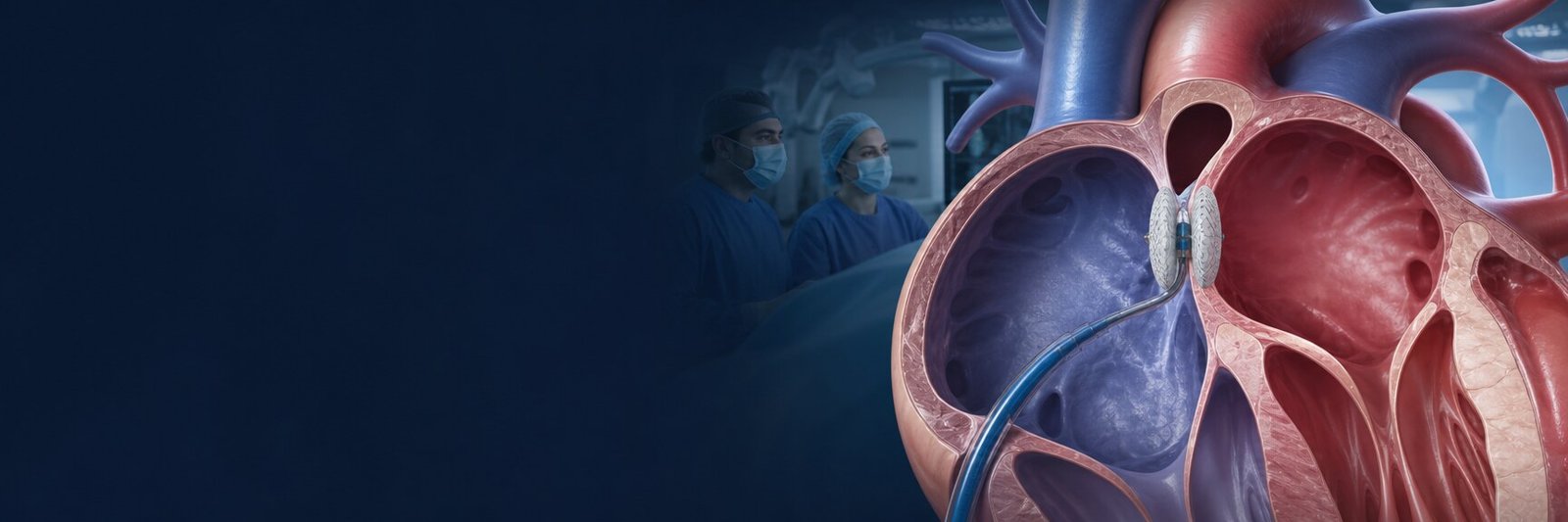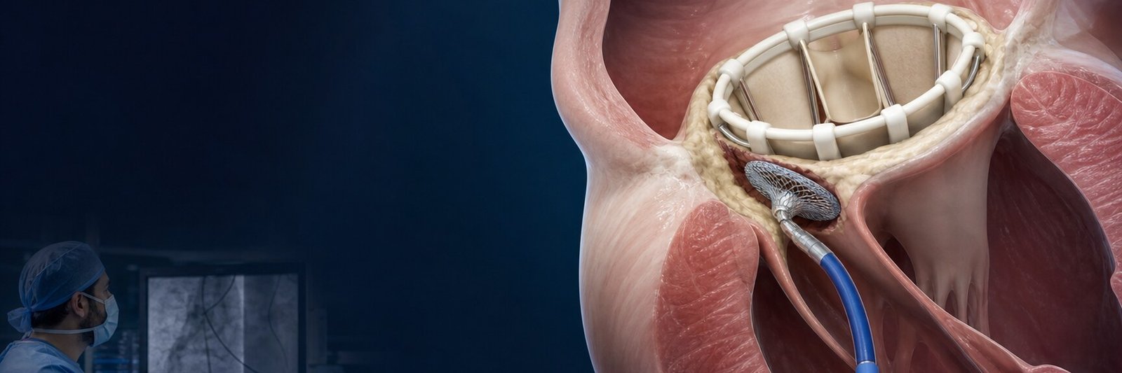
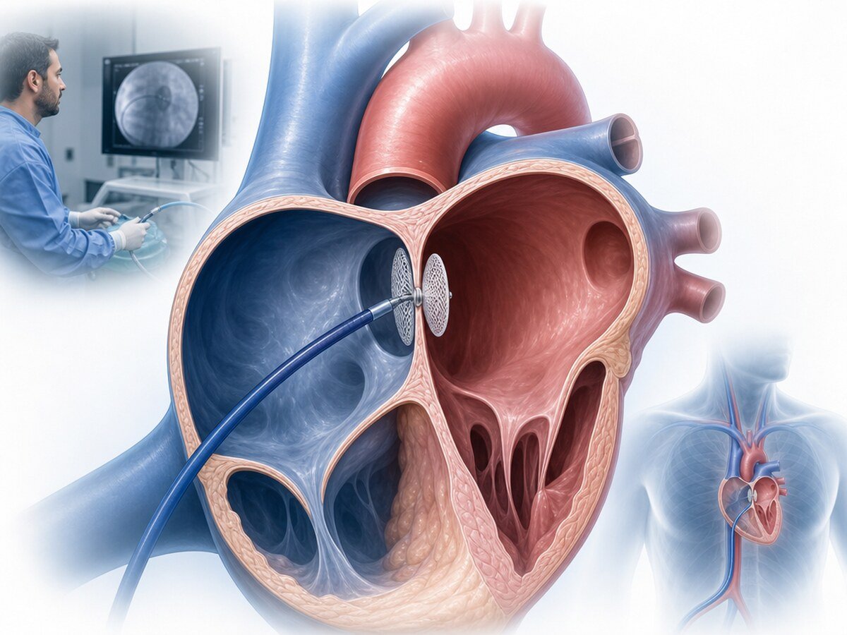
PFO closure is placement of a closure device across a small passage between the heart’s upper chambers that did not seal completely after birth. The catheter is usually advanced from a femoral vein. Finding a patent foramen ovale (PFO) is not, by itself, a reason to close it. The decision follows a review of other possible stroke causes and of whether this PFO is related to the event that occurred.[1,3,4]
What is a PFO?
A patent foramen ovale is a small opening in the wall between the right and left atria that was needed before birth and did not close completely afterwards. Before birth, some blood normally bypasses the lungs through this passage. After birth the lungs take over and the opening usually seals; in some people a flap-like passage remains.[4,16]
A PFO is common and may be present in about one in four adults. Most people never have symptoms. Seeing a PFO on echocardiography is not, by itself, a disease diagnosis or an explanation for a previous stroke.[4,16]
A PFO is not the same as a large hole in the heart. Continuous wide flow is usually absent. Right-to-left passage can become clearer with coughing, straining or a specialised ultrasound study.
Why does a PFO remain open?
A PFO is usually a developmental feature, not a tear that forms later. Clinical importance depends on how much blood crosses, the structure of the atrial septum and whether the timing and mechanism fit the person’s event.[5,11]
What is PFO closure?
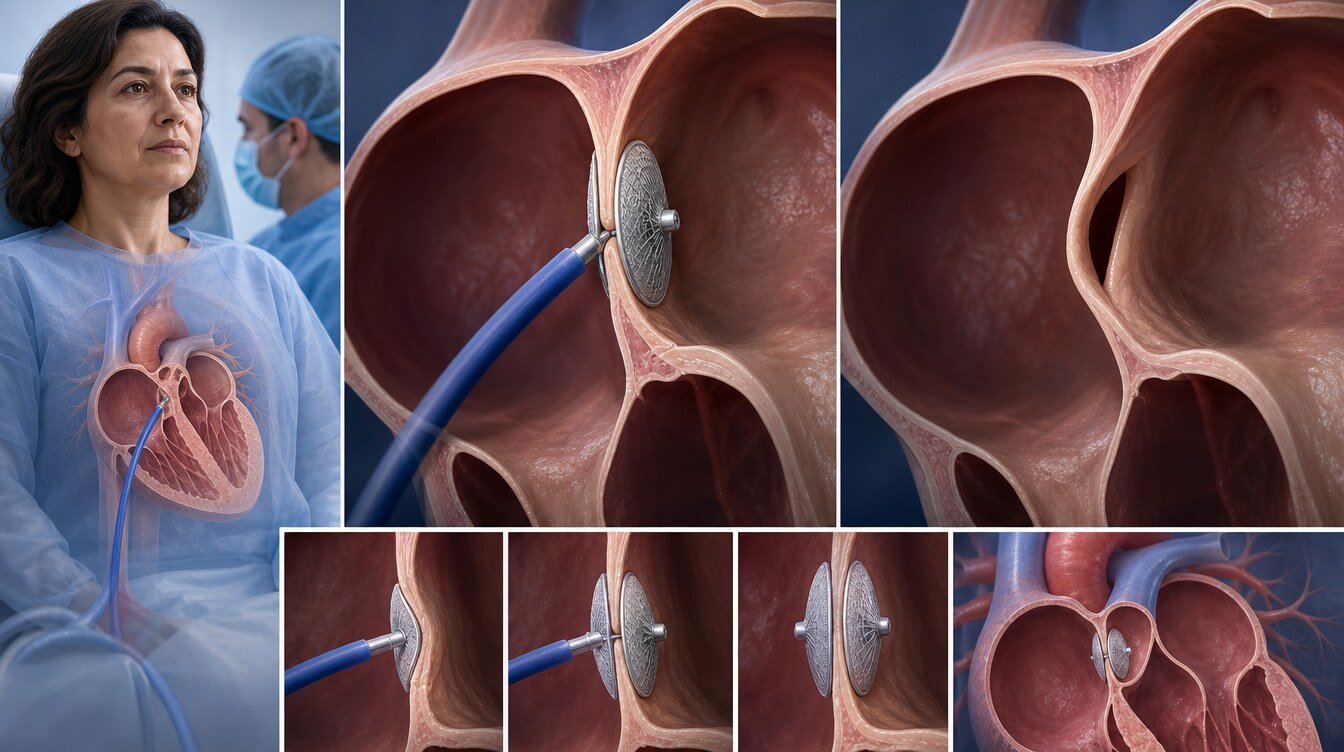
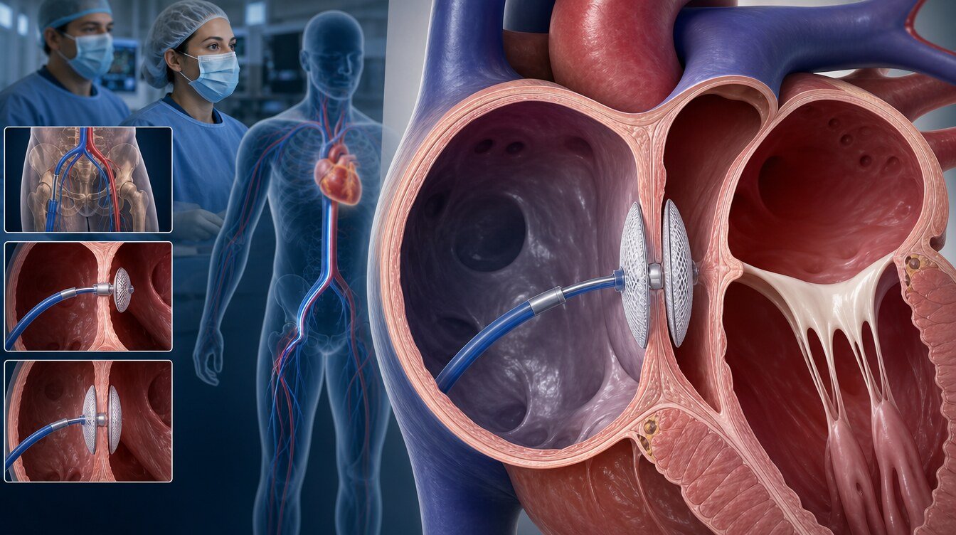
PFO closure is one of the transcatheter procedures in structural heart interventions. A thin, flexible catheter carries a device that opens on both sides of the passage and grasps the septal wall, aiming to reduce or stop right-to-left flow.[3,11]
This is not open-heart surgery. Avoiding a chest incision does not make the procedure simple, risk-free or suitable for everyone. ASD closure, LAA closure and PVL closure answer different anatomical and clinical questions and are not interchangeable.
How is a PFO related to ischaemic stroke?
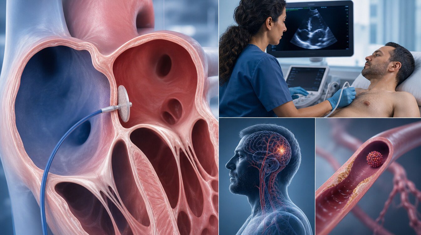
An ischaemic stroke occurs when a brain artery is blocked by clot or another material. One possible PFO-related mechanism is paradoxical embolism: a clot from the veins may cross from the right atrium to the left atrium through the PFO instead of travelling to the lungs, then reach arteries that supply the brain.[3,10]
Finding a PFO does not prove that the stroke came from the PFO. Two questions are asked:
- Is there a PFO? Imaging looks for the passage.
- Is this PFO related to the stroke? Other causes and the likelihood of a causal role are reviewed.
Small-vessel disease, atherosclerosis, carotid disease, cardiac thrombus or arrhythmia may be a stronger explanation than the PFO. Atrial fibrillation, which can be intermittent and easy to miss, should be investigated.[4,15]
A large right-to-left shunt and an atrial septal aneurysm can raise the chance that the PFO is related to the stroke. Benefit of closure has been clearer in selected patients with both features. That is not an automatic indication for every large shunt or every aneurysm.[3,12]
Should every PFO be closed?
No. Because PFO is common, most people with a PFO do not need closure. Closure is not routinely advised when the PFO is an incidental finding, when there is no stroke history, or when another cause of stroke is present.[3,4]
Even after an ischaemic stroke, a PFO is not an automatic reason to close. Imaging of the stroke, vessels, heart rhythm, other cardiac sources and vascular risk factors come first. If another mechanism is more likely, closing the PFO alone will not remove that mechanism. The American Academy of Neurology recommends appropriate, thorough exclusion of alternative stroke causes before closure is considered.[4]
How selected trial evidence is read is also discussed in How does PFO closure reduce stroke risk?.
Who may be considered?
PFO closure may be assessed in people who:
- are usually between 18 and 60 years of age
- have an imaging-confirmed ischaemic stroke
- have no other sufficient cause after adequate investigation
- have a stroke pattern that appears embolic
- have higher-risk structural or functional PFO features
Even then the decision is individual. CLOSE, REDUCE, RESPECT and DEFENSE-PFO showed that, in selected patients with cryptogenic stroke, closure can reduce recurrent stroke compared with medication alone. Those trials mainly enrolled people aged 60 or younger, so the results are not applied automatically to every older adult.[3,6–9,15]
When is closure not a routine option?
Closure is usually not a routine choice when:
- a PFO is found incidentally and there is no stroke history
- another explanation for stroke is stronger than the PFO
- “cryptogenic” is declared before adequate rhythm investigation
- the request is based on migraine alone
- expected benefit is uncertain and procedure or bleeding risk is judged high for that person
Unsuitability is not decided by one age cut-off or one imaging number. Neurology and cardiology interpret alternative causes, PFO features and preferences together.[1,3,4,14]

What is the RoPE score?
The RoPE score (Risk of Paradoxical Embolism) uses age, vascular risk factors and stroke-imaging features to help estimate how likely the PFO is related to the stroke. A lower score may suggest an incidental PFO; a higher score may suggest a more plausible causal role.[15,22]
The score is not a diagnostic test that decides closure on its own. It does not replace imaging, rhythm investigation or the clinical history.
What is the PASCAL classification?
PASCAL combines the RoPE score with higher-risk anatomical PFO features to support judgement about whether the PFO is a probable, possible or unlikely contributor. It also does not replace clinical assessment, imaging and other tests. The European Stroke Organisation notes that PASCAL may be used in selected patients.[3]
What is a large right-to-left shunt?
A right-to-left shunt is blood crossing from the right atrium to the left atrium through the PFO. “Large shunt” is usually defined by how many bubbles or how much flow reach the left heart during a bubble study. Definitions vary by centre and method.
A large shunt is one feature that can make an incidental PFO less likely. It does not decide closure by itself; other stroke causes and the whole clinical picture still matter.[3,12]
What is an atrial septal aneurysm?
An atrial septal aneurysm is a septum that is more mobile or ballooning than usual. When it accompanies a PFO, some analyses have shown a clearer link with recurrent stroke and with benefit from closure. An aneurysm is not an automatic procedural indication either.[12]
How are alternative stroke causes excluded?
Before closure is considered, other explanations are investigated. The work-up is individual; there is no single universal list. Common elements include:[4,15]
- CT or MRI of the brain to define the stroke
- imaging of brain and neck vessels
- assessment of the carotid and other arteries
- blood pressure, diabetes, cholesterol, smoking and other vascular risks
- search for thrombus or another cardiac embolic source
- clotting tests when they are clinically justified
If another cause is more likely, PFO closure does not remove that cause.
How is atrial fibrillation investigated?
Atrial fibrillation is an irregular, often rapid rhythm. A short recording may miss it. Electrocardiography and, when appropriate, more prolonged rhythm monitoring may therefore be planned.[4,15]
Declaring a stroke “cryptogenic, therefore close the PFO” before rhythm investigation is incomplete. If atrial fibrillation is found, prevention may change and anticoagulation may be needed.
How is a patient assessed before the procedure?
Both the presence of a PFO and the features of the shunt are reviewed. Transthoracic echocardiography looks from the chest wall. Transoesophageal echocardiography, used in some patients, shows PFO anatomy, shunt size, an atrial septal aneurysm and other cardiac sources in more detail.[3]
Brain and vessel imaging, electrocardiography, rhythm monitoring of appropriate duration, bleeding risk, kidney function, allergies and current medicines are also reviewed. Which tests are needed depends on previous results and the history. The question is not only whether a device can be placed, but whether the procedure is likely to help that person.[15]
Cardiology and neurology usually decide together.
How is the procedure performed?
Details vary with anatomy, device and centre protocol. In general:[11]
- The patient is reviewed again; the sedation or anaesthesia plan is explained.
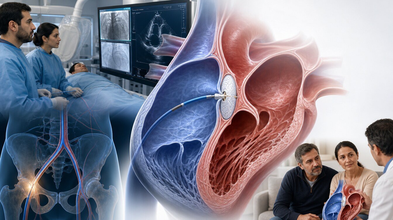
- Venous access is usually obtained in the groin.
- The catheter is advanced to the right atrium.
- Under imaging, the catheter crosses the PFO into the left atrium.
- One part of the device opens on the left side of the septum and the other on the right, grasping both faces.
- Position, wall apposition and residual flow are checked; the device can be repositioned if needed.
- The catheter is removed, pressure is applied at the access site and a period of observation follows.
Procedure duration is not the same for everyone. Ultrasound and X-ray imaging may be used; the form of anaesthesia varies. A web page should not be used to derive one universal duration, fasting time or discharge day.
What is a PFO closure device?
A PFO closure device is a small implant that opens on both sides of the passage and holds the septal tissue. The aim is to reduce or stop right-to-left flow. Position and apposition are checked with imaging during placement.[3,13]
Heart tissue is expected to cover the device over time. Antiplatelet medicines, which reduce platelet clumping, may be used to lower the chance of clot on the device surface. Drug type and duration vary; one prescription should not be copied from a website.[3,6]
PFO anatomy is not uniform. Device choice depends on pre-procedure imaging and intra-procedural checks. This page does not compare brands or give a technical recipe.
What are the risks and specific complications?
PFO closure is an invasive procedure whose complication risk is not zero. Individual risk depends on anatomy, device, other illness and how follow-up is done.[11,13]
Risks that are discussed include:
- groin bleeding, bruising, pain, venous thrombosis or, rarely, vessel injury
- palpitations or new atrial fibrillation
- clot on the device
- device migration or the need to reposition
- residual shunt after closure
- rarely, cardiac injury, perforation or fluid around the heart
- contrast allergy or a temporary change in kidney function
- infection at the access site or, more rarely, related to the device
A meta-analysis of randomised trials reported major procedural complications in 2.40 percent of patients. CLOSE reported procedure-related complications in 5.9 percent. Those figures belong to study definitions; they are not a personal risk calculation.[6,13]
New atrial fibrillation
New atrial fibrillation can occur after the procedure. In one meta-analysis of controlled studies it was reported in 5.1 percent after closure and 1.6 percent with medical therapy. In CLOSE the figures were 4.6 percent after closure and 0.9 percent with antiplatelet therapy alone. More intensive monitoring can detect more episodes. Some events appear early and settle; if AF is found, follow-up is individual. Published rates are not a personal estimate.[6,17,19]
Residual shunt
Some flow across the PFO can remain after closure. Echocardiography, and a bubble study when needed, assess this. Residual shunt has been linked in some studies with recurrent stroke risk; management depends on the amount of shunt and the clinical picture. Residual flow does not automatically mean the procedure “failed” or that all protection is lost.[18]
What medicines are used afterwards?
Most protocols use antiplatelet therapy for a period to lower the chance of clot on the device. Some studies used two antiplatelet drugs and then one. Duration and choice vary with protocol, device and bleeding risk. There is no single regimen for everyone.[3,6]
Anticoagulants that suppress the clotting system are not used routinely and long term for a PFO alone. They may be needed if atrial fibrillation, venous thrombosis or another medical reason is present. The European Stroke Organisation advises against long-term anticoagulation after PFO-associated stroke when no other indication exists.[3]
Medicines should not be stopped or changed without medical advice. Dental work, an operation or a new drug should be discussed with the team that knows about the closure.
How is follow-up arranged?
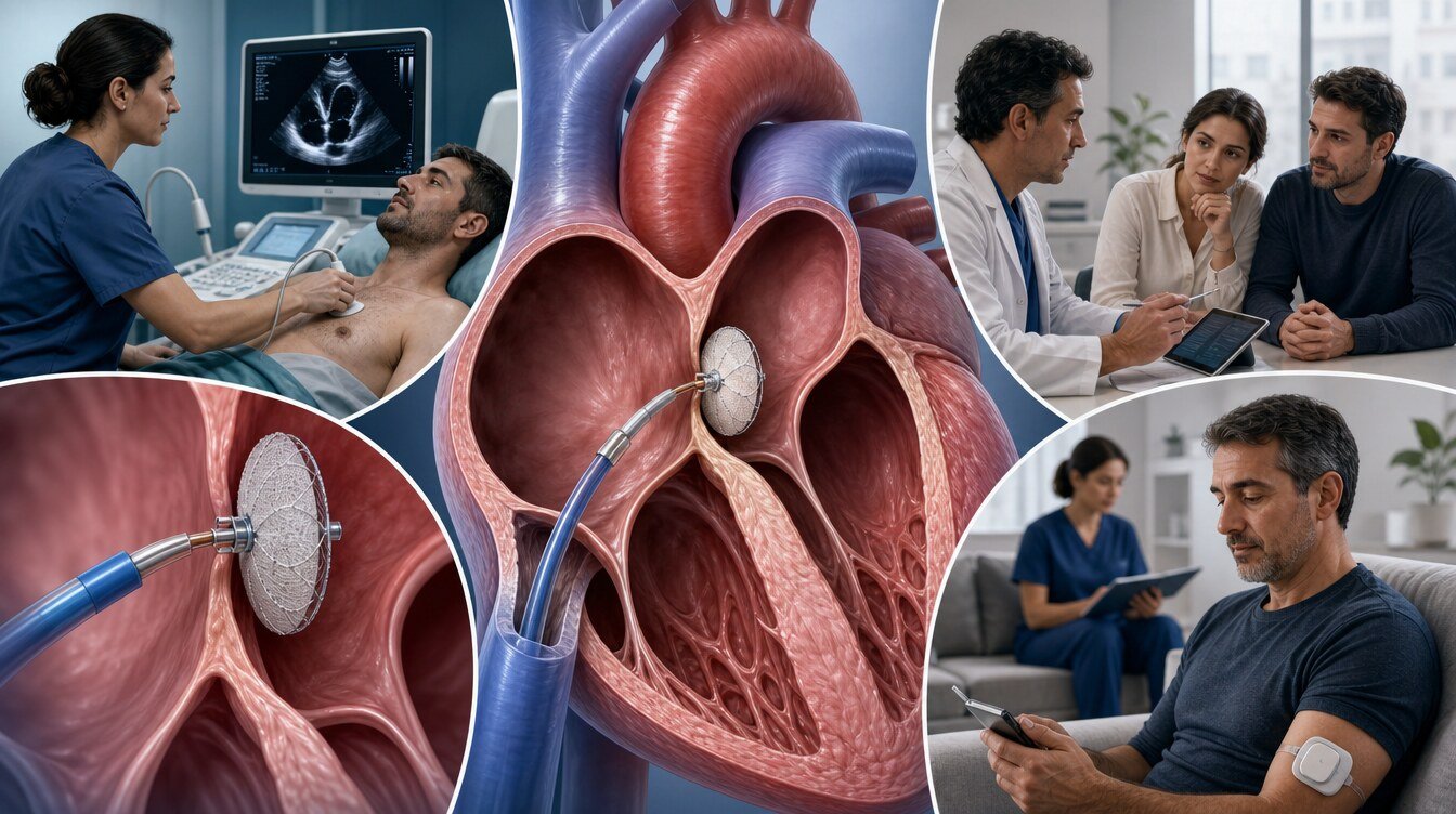
Follow-up checks device position, wall apposition, residual flow and heart rhythm. Echocardiography may be used; a bubble study can look for remaining right-to-left passage. Timing depends on the device, procedural findings and the clinical course.[3]
Observation time, same-day or short hospital stay and return to daily life vary. Mild groin pain or bruising can occur in the first days. Rapidly enlarging swelling, ongoing bleeding, severe pain, fainting, breathlessness, chest pain or marked palpitations need medical review.[17,19]
Sudden facial droop, arm or leg weakness, speech difficulty or sudden visual loss may be stroke symptoms. In Türkiye, call 112.
What changes after age 60?
Most randomised trials enrolled people aged 18–60. For patients aged 60 or older who have already had a PFO-associated stroke, guidelines read the evidence differently.
SCAI 2022 gives a conditional recommendation for PFO closure versus long-term antiplatelet therapy in that group. Certainty of evidence is very low. It is also reasonable for patients who value avoiding procedural risk more than an uncertain benefit to decline closure.[1,2]
ESO 2024 states that the evidence is not sufficient for an evidence-based recommendation after age 60.[3]
That difference does not mean “routine closure after 60” or “never close after 60.” The decision follows thorough exclusion of atrial fibrillation and vascular disease, then a discussion of individual risks and preferences.
PFO and migraine
Observational studies have reported a link between PFO and migraine. Closure is not an accepted routine indication for treating migraine. In the PREMIUM trial, closure did not meet the primary migraine-response end point in people with frequent migraine. Migraine alone is not a reason to close a PFO.[14,20]
Migraine symptoms may change in some people who undergo closure for a PFO-related stroke. That possible change is not the purpose or a guarantee of the procedure. Migraine care is planned through neurological assessment.
What happens if the PFO is not closed?
Most people have no problem if a PFO is left open, because the finding is common and most people live with it.[4]
After a cryptogenic ischaemic stroke, when no other cause is found and the PFO may be related to the event, a choice is made between closure and medical therapy. If closure is not performed, antiplatelet therapy or, in some situations, anticoagulation may be considered; benefit and bleeding risk are individual.[3,4]
Not closing does not mean another stroke will certainly occur. Closing also does not prevent every future stroke, because other mechanisms can still operate. Randomised-trial benefit has been clearest in selected patients with cryptogenic stroke who were younger than 60.[3,7,8]
Frequently asked questions
No. It is usually a catheter-based procedure through a femoral vein, not an open-chest operation. It is still an invasive intervention that needs vascular access, imaging, device placement and follow-up.[11]
Yes. The randomised trials mainly enrolled people aged 18–60. SCAI 2022 gives a conditional recommendation for closure versus long-term antiplatelet therapy after a prior PFO-associated stroke at age 60 or older, with very low certainty. ESO 2024 says the evidence is insufficient for an evidence-based recommendation in this age group. That is not a routine-closure rule.[1,3]
The RoPE score helps estimate how likely the PFO is related to the stroke. It is not a stand-alone test that decides closure.[15]
A small residual passage can persist. Follow-up echocardiography measures it; the amount of shunt and the clinical picture are interpreted together.[18]
No. Closure is not a routine treatment for migraine. A randomised trial did not meet its primary migraine-response end point.[14]
Assessment and decision
The PFO closure decision does not rest on the sentence “a PFO is present.” Stroke features, exclusion of other causes, atrial-fibrillation investigation, shunt and atrial septal aneurysm, supportive tools such as RoPE and PASCAL, the age-related guideline difference and the person’s preferences are read together.[1,3,4]
Literature
References
Kavinsky CJ, et al. SCAI Guidelines for the Management of Patent Foramen Ovale. Journal of the Society for Cardiovascular Angiography & Interventions. 2022. doi:DOI: 10.1016/j.jscai.2022.100039.
Goldsweig AM, et al. SCAI Technical Review on Management of Patent Foramen Ovale. Journal of the Society for Cardiovascular Angiography & Interventions. 2022. doi:DOI: 10.1016/j.jscai.2022.100040.
Caso V, et al. European Stroke Organisation (ESO) Guidelines on the diagnosis and management of patent foramen ovale (PFO) after stroke. European Stroke Journal. 2024. doi:DOI: 10.1177/23969873241247978.
Messé SR, et al. Practice advisory update summary: Patent foramen ovale and secondary stroke prevention. Neurology. 2020. doi:DOI: 10.1212/WNL.0000000000009443. PMID 32350064.
Sposato LA, et al. Patent Foramen Ovale Management for Secondary Stroke Prevention: State-of-the-Art Appraisal of Current Evidence. Stroke. 2024. doi:DOI: 10.1161/STROKEAHA.123.040546.
Mas JL, et al. Patent Foramen Ovale Closure or Anticoagulation vs. Antiplatelets after Stroke. N Engl J Med. 2017. doi:DOI: 10.1056/NEJMoa1705915. PMID 28902580.
Søndergaard L, et al. Patent Foramen Ovale Closure or Antiplatelet Therapy for Cryptogenic Stroke. N Engl J Med. 2017. doi:DOI: 10.1056/NEJMoa1707404. PMID 28902593.
Saver JL, et al. Long-Term Outcomes of Patent Foramen Ovale Closure or Medical Therapy after Stroke. N Engl J Med. 2017. doi:DOI: 10.1056/NEJMoa1610057. PMID 28902590.
Lee PH, et al. Cryptogenic Stroke and High-Risk Patent Foramen Ovale. J Am Coll Cardiol. 2018. doi:DOI: 10.1016/j.jacc.2018.02.046. PMID 29699609.
Elgendy AY, et al. Proposal for Updated Nomenclature and Classification of Potential Causative Mechanism in Patent Foramen Ovale–Associated Stroke. JAMA Neurology. 2020. doi:DOI: 10.1001/jamaneurol.2020.0458. PMID 32227141.
Alkhouli M, et al. Patent foramen ovale closure for secondary stroke prevention. European Heart Journal. 2019. doi:DOI: 10.1093/eurheartj/ehz157.
Turc G, et al. Atrial Septal Aneurysm, Shunt Size, and Recurrent Stroke Risk in Patients With Patent Foramen Ovale. J Am Coll Cardiol. 2020. doi:DOI: 10.1016/j.jacc.2020.02.068. PMID 32381164.
Turc G, et al. Closure, Anticoagulation, or Antiplatelet Therapy for Cryptogenic Stroke With Patent Foramen Ovale. Journal of the American Heart Association. 2018. doi:DOI: 10.1161/JAHA.117.008356. PMID 29514809.
Tobis JM, et al. Percutaneous Closure of Patent Foramen Ovale in Patients With Migraine. J Am Coll Cardiol. 2017. doi:DOI: 10.1016/j.jacc.2017.09.1105. PMID 29150278.
Elzanaty A, et al. Patent foramen ovale closure in the management of cryptogenic stroke: a review of current literature and guideline statements. Current Medical Research and Opinion. 2021. doi:DOI: 10.1080/03007995.2021.1876648.
Lucà F, et al. Patent Foramen Ovale and Cryptogenic Stroke: Integrated Management. Journal of Clinical Medicine. 2023. doi:DOI: 10.3390/jcm12051952.
Apostolos A, et al. Atrial Fibrillation After Patent Foramen Ovale Closure: Incidence, Pathophysiology, and Management. Journal of the American Heart Association. 2024. doi:DOI: 10.1161/JAHA.124.034249.
Rhoades R, et al. Secondary stroke prevention in patients with patent foramen ovale. Current Opinion in Hematology. 2021. doi:DOI: 10.1097/MOH.0000000000000672.
Jurczyk D, et al. New-onset atrial fibrillation after percutaneous patent foramen ovale closure: a meta-analysis. Clinical Research in Cardiology. 2023. doi:DOI: 10.1007/s00392-023-02263-8.
Mojadidi MK, et al. Pooled Analysis of PFO Occluder Device Trials in Patients With PFO and Migraine. J Am Coll Cardiol. 2021. doi:DOI: 10.1016/j.jacc.2020.11.068.
Müller F, et al. PFO closure in ischemic stroke: insights from a single-center real-world cohort. Frontiers in Neurology. 2026. doi:DOI: 10.3389/fneur.2026.1792925.
Rahman S, et al. Association between RoPE score and PFO grading on bubble echocardiography in cryptogenic stroke patients: a retrospective cohort study. Frontiers in Stroke. 2025. doi:DOI: 10.3389/fstro.2025.1676220.
This content is for general information only and does not replace personalized medical advice. In emergencies call local emergency services.
Continue