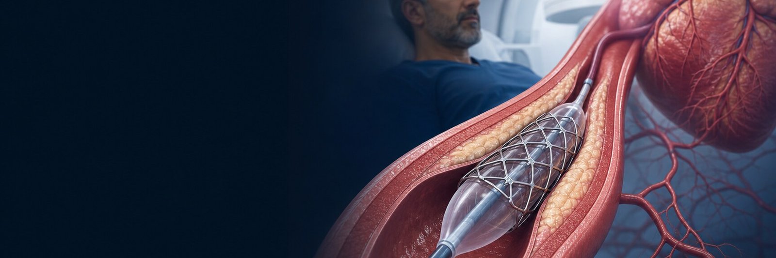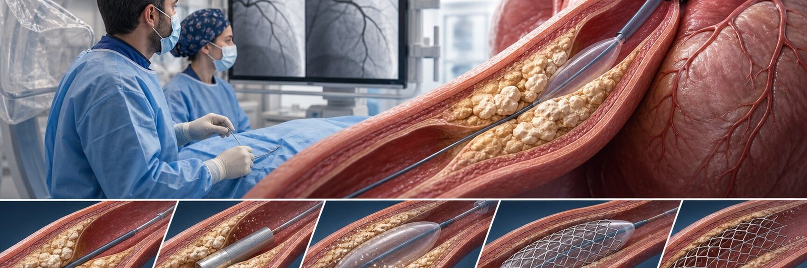
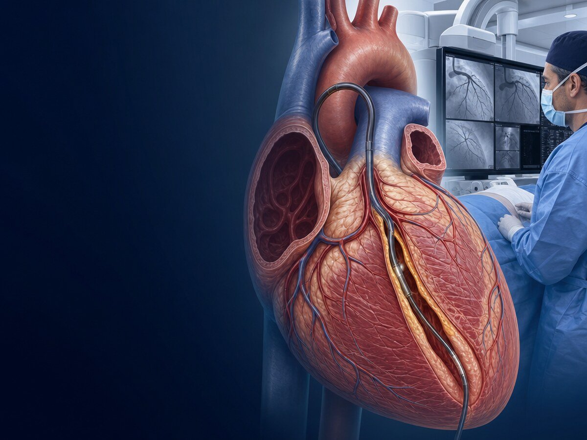
Coronary angiography is an invasive diagnostic method that images the inside of the coronary arteries that supply the heart, using contrast dye and X-rays. It is often called a “heart angiogram” or simply “angiography.”
The procedure helps assess whether there is narrowing or blockage in a coronary artery, and if so, where it is and what the anatomy looks like. However, having angiography does not always mean a stent will be placed. In some patients imaging alone is enough; some narrowings need additional measurement; in others medication, stent or bypass options are considered together.
What does coronary angiography show?
The coronary arteries carry oxygen and nutrients to the heart muscle. Narrowing from atherosclerotic plaque can reduce blood flow. Coronary angiography can help assess:
- Where narrowing or blockage is located,
- Which segment of the vessel is affected,
- Whether more than one artery is involved,
- General features relevant to stenting or surgery,
- Previously placed stents and bypass grafts.
Angiography shows anatomy. Moderate-appearing narrowings may not always be judged from the image alone as to whether they truly limit blood flow. In such cases FFR or iFR measurement or intravascular imaging may be added.[1,8–10]
Who needs coronary angiography?
The decision is not based on a single symptom or test. Current guidelines especially consider angiography when:[1–3]
- Heart attack or high-risk acute coronary syndrome is suspected,
- Chest pain or breathlessness significantly affects daily life despite medication,
- Symptoms suggest coronary disease at low exercise levels,
- Stress testing, stress imaging or coronary CT angiography shows high-risk findings,
- Non-invasive tests are inconclusive but coronary disease remains likely,
- Reduced heart pump function raises suspicion of widespread coronary disease,
- Prior stent or bypass and new findings suggest recurrent vessel problems,
- Coronary arteries must be assessed before certain valve or cardiac surgery.
Having one of these features does not mean angiography is mandatory for everyone. Age, kidney function, bleeding risk, other illnesses, symptom pattern and prior work-up all matter.
When is emergency angiography needed?
High-risk presentations such as ECG changes suggesting heart attack, ongoing or recurrent chest pain, shock, life-threatening rhythm problems or heart failure need urgent evaluation. The aim is not only diagnosis but, when appropriate, treating a blocked artery in the same session.[2,3]
Does every stable chest pain need immediate angiography?
No. In stable patients with low or intermediate likelihood of coronary artery disease, coronary CT angiography or an appropriate functional stress test may be first. Invasive angiography is more strongly considered with high pre-test probability, severe or refractory symptoms, high-risk test results or persistent uncertainty.[1,4]
How is coronary angiography performed?
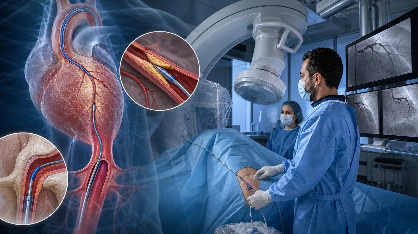
The procedure is done in a catheter laboratory under sterile conditions. The usual steps are:
- Preparation and monitoring: Blood pressure, pulse and rhythm are monitored; intravenous access is placed and relevant labs and history are checked.
- Local anaesthesia: The radial artery at the wrist or femoral artery in the groin is numbed. Most patients remain awake.
- Catheter advancement: A thin sheath and catheter are advanced to the coronary ostia.
- Imaging: Contrast is injected and short X-ray runs are recorded. A brief warm flush may be felt.
- Additional assessment if needed: FFR/iFR for intermediate lesions; IVUS or OCT for detailed vessel or stent assessment.
- Completion: Catheter and sheath are removed; wrist compression or groin closure is applied.
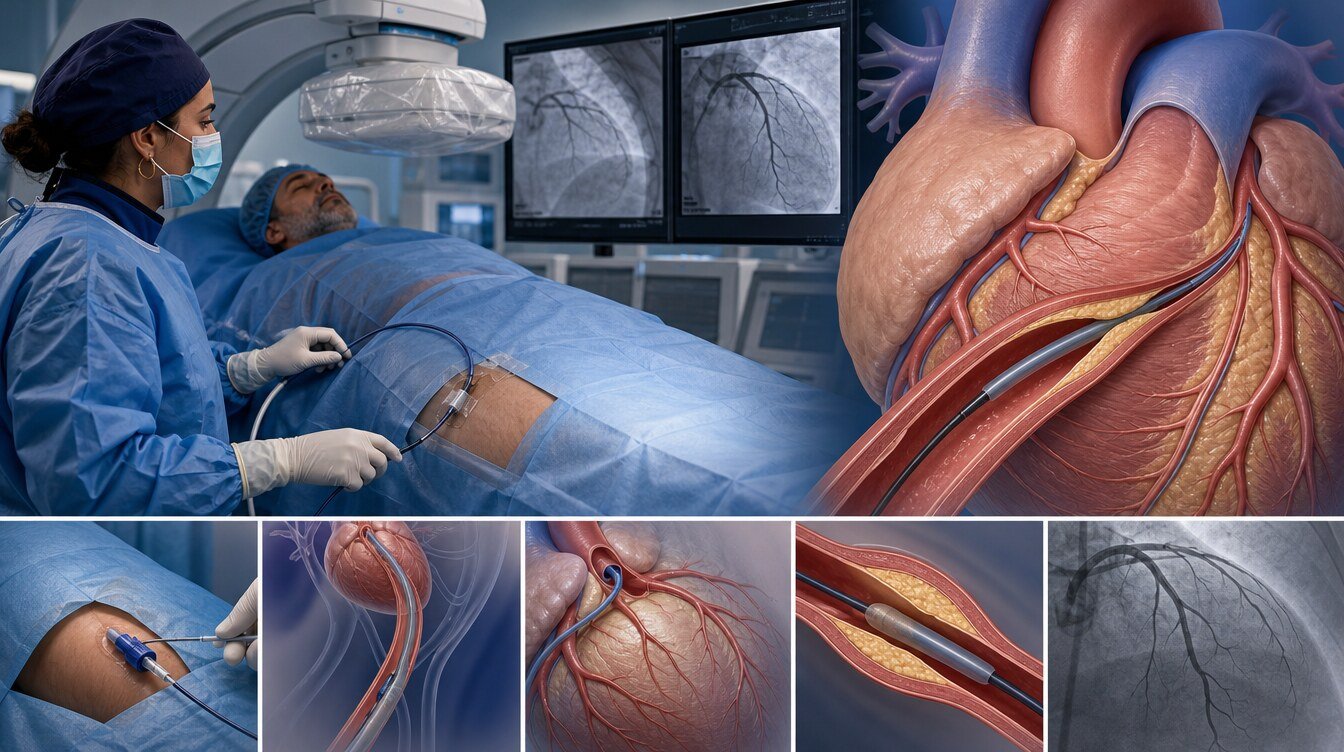
Diagnostic imaging itself is often relatively short, but total hospital time includes preparation, possible add-on tests, closure and observation. Timing also depends on prior bypass or stents and whether treatment is done in the same session.
Radial (wrist) or femoral (groin) access?
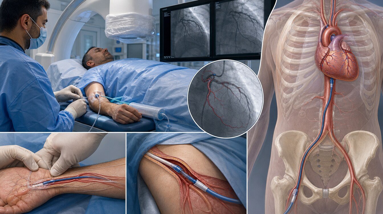
Coronary angiography can be done through the radial artery at the wrist or the femoral artery in the groin.
Radial (wrist) angiography
In suitable patients, radial access is the standard approach supported by guidelines and randomized trials. It reduces bleeding and access-site complications, especially in acute coronary syndromes, and often allows earlier mobilization.[1–3,5–7]
Femoral (groin) angiography
Femoral access may be more appropriate when the wrist vessel is small or unusable, larger equipment is needed, or anatomy and planned complexity require it.
| Feature | Wrist (radial) | Groin (femoral) |
|---|---|---|
| Access-site bleeding | Generally lower | May be relatively higher |
| Mobilization after procedure | Often earlier | May require lying flat for a period |
| Technical suitability | Not possible in every patient | May be needed for some complex cases |
| Preference | Often first choice when suitable | Important option when clinically indicated |
Why might FFR or iFR be needed?
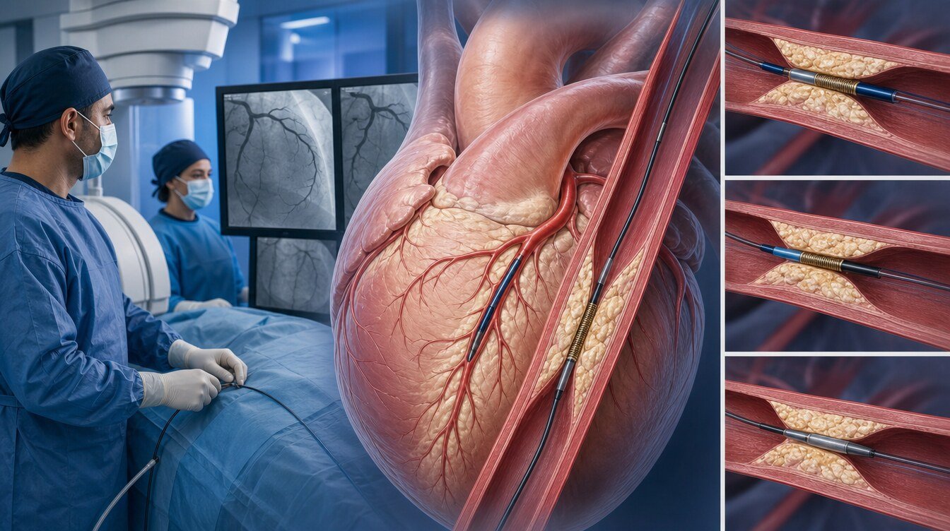
The percentage narrowing on angiography does not always show how much blood flow is affected. For intermediate lesions, FFR uses a pressure wire (often with a vasodilator); iFR assesses pressure difference in a specific part of the cardiac cycle and usually needs no extra vasodilator. European guidance recommends functional confirmation with FFR or iFR when intervention is considered.[1] Major trials show similar outcomes with iFR- versus FFR-guided decisions.[9,10]
Is a stent always placed during angiography?
No. Coronary angiography is diagnostic; stenting is one treatment option when appropriate. Outcomes may include no significant stenosis, medical therapy, FFR/iFR or intravascular imaging, same- or later-session stent, or surgical opinion for complex disease. Outside emergencies, same-session stenting and alternatives should be discussed beforehand. In complex disease, the decision weighs clinical status, anatomy, comorbidities and patient preference.
Preparation before coronary angiography
Follow the hospital’s personal instructions. Tell the team about all medications (including blood thinners and diabetes drugs), kidney disease, prior contrast reactions, bleeding disorders, pregnancy possibility, and prior angiography, stent, bypass or surgery. Do not stop prescribed medicines without medical advice.
What if kidney disease is present?
Contrast can affect kidney function in some patients. Risk rises with existing kidney disease, advanced age, diabetes, heart failure, dehydration and higher contrast volume. The main approach is pre-assessment, appropriate IV fluid planning when needed, and limiting contrast volume.[11–13] The same fluid or drug protocol is not applied to everyone. The large PRESERVE trial did not show superiority of sodium bicarbonate over normal saline, or of acetylcysteine over placebo.[12]
What are the risks of coronary angiography?
Most complications are manageable access-site issues, but angiography is invasive and not risk-free. Possible risks include access-site pain, bruising or bleeding; vessel injury; contrast reaction; kidney injury; rhythm changes; coronary artery injury; and very rarely heart attack, stroke, emergency surgery or death. The 2024 ESC chronic coronary syndrome guideline reports pooled major complication rates around 0.5–2% with femoral diagnostic catheterization and around 0.1–0.2% for death, MI or stroke with radial access.[1] Individual risk varies.
Pre-procedure assessment, radial access when suitable, bleeding-risk review, kidney protection planning and limiting contrast volume are key steps to reduce risk.[1–3,7,11]
After angiography — what to watch for
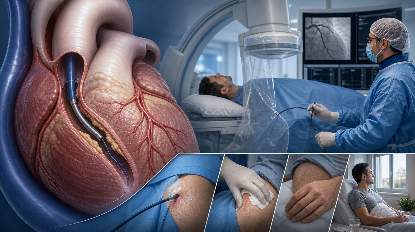
Pulse, blood pressure, rhythm and the access site are monitored. Discharge timing depends on whether only diagnostics were done, access route, additional treatment and overall condition. Seek help promptly for bleeding that does not stop with pressure, rapidly enlarging swelling, cold or weak limb, new or worsening chest pain, severe breathlessness, fainting, allergic-type symptoms or reduced urine output.
Coronary angiography vs coronary CT angiography
| Feature | Invasive coronary angiography | Coronary CT angiography |
|---|---|---|
| Approach | Catheter via artery | IV contrast and CT scan |
| Main goal | Direct vessel imaging; same-session treatment if needed | Non-invasive anatomy and rule-out |
| Same-session stent | Possible when appropriate | Not possible |
| Typical use | High risk, severe symptoms, emergencies, selected uncertainty | Many stable low–intermediate probability patients |
Frequently asked questions
The access site is numbed with local anaesthetic. Brief burning, pressure or a warm flush with contrast may occur.
Diagnostic coronary angiography is usually done with local anaesthesia while you are awake. Sedation may be given if needed.
Diagnostic imaging is often short, but vessel anatomy, add-on tests and same-session treatment change timing. Total hospital stay is longer than the imaging itself.
No. Radial access is often preferred when suitable, but groin access may be chosen because of vessel anatomy or technical needs.
Not always. Significance, symptoms, FFR/iFR, anatomy and bypass options are assessed together.
Not always. Microvascular dysfunction or coronary spasm may still be present; non-cardiac causes are also possible.[1]
This depends on access route and whether only diagnostics or stenting was done. Follow the personal advice on your discharge instructions.
Assessment and decision
The decision should rest on whether the result will change management, not only on whether narrowing exists. Test sequence, access route, kidney and bleeding risk, and same-session add-on procedures are discussed during evaluation.
Literature
References
Vrints C, Andreotti F, Koskinas KC, et al. 2024 ESC Guidelines for the management of chronic coronary syndromes. European Heart Journal. 2024;45(36):3415–3537. DOI: 10.1093/eurheartj/ehae177
Rao SV, O'Donoghue ML, Ruel M, et al. 2025 ACC/AHA/ACEP/NAEMSP/SCAI Guideline for the Management of Patients With Acute Coronary Syndromes. Circulation. 2025. DOI: 10.1161/CIR.0000000000001309
Byrne RA, Rossello X, Coughlan JJ, et al. 2023 ESC Guidelines for the management of acute coronary syndromes. European Heart Journal. 2023;44(38):3720–3826. DOI: 10.1093/eurheartj/ehad191
Gulati M, Levy PD, Mukherjee D, et al. 2021 AHA/ACC/ASE/CHEST/SAEM/SCCT/SCMR Guideline for the Evaluation and Diagnosis of Chest Pain. Circulation. 2021;144:e368–e454. DOI: 10.1161/CIR.0000000000001029
Jolly SS, Yusuf S, Cairns J, et al. RIVAL. The Lancet. 2011;377(9775):1409–1420. DOI: 10.1016/S0140-6736(11)60404-2
Valgimigli M, Gagnor A, Calabró P, et al. MATRIX. The Lancet. 2015;385(9986):2465–2476. DOI: 10.1016/S0140-6736(15)60292-6
Bajraktari G, Rexhaj Z, Elezi S, et al. Radial Access Meta-Analysis. Journal of Clinical Medicine. 2021;10(10):2163. DOI: 10.3390/jcm10102163
Koo BK, Hu X, Kang J, et al. FFR or IVUS to Guide PCI. NEJM. 2022;387:779–789. DOI: 10.1056/NEJMoa2201546
Götberg M, Christiansen EH, et al. iFR vs FFR to Guide PCI. NEJM. 2017;376:1813–1823. DOI: 10.1056/NEJMoa1616540
Davies JE, Sen S, et al. iFR or FFR in PCI. NEJM. 2017;376:1824–1834. DOI: 10.1056/NEJMoa1700445
Lawton JS, et al. 2021 ACC/AHA/SCAI Revascularization Guideline. Circulation. 2022;145:e18–e114. DOI: 10.1161/CIR.0000000000001038
Weisbord SD, et al. PRESERVE. NEJM. 2018;378:603–614. DOI: 10.1056/NEJMoa1710933
van der Molen AJ, et al. Post-contrast AKI — Part 2. European Radiology. 2018;28:2856–2869. DOI: 10.1007/s00330-017-5247-4
Continue