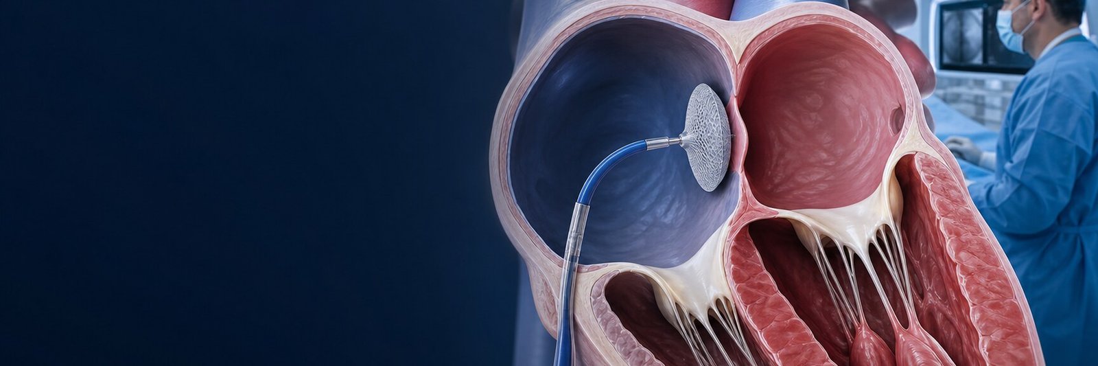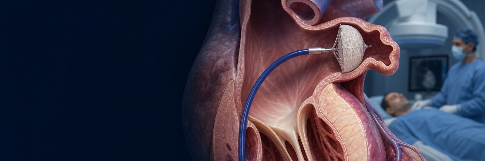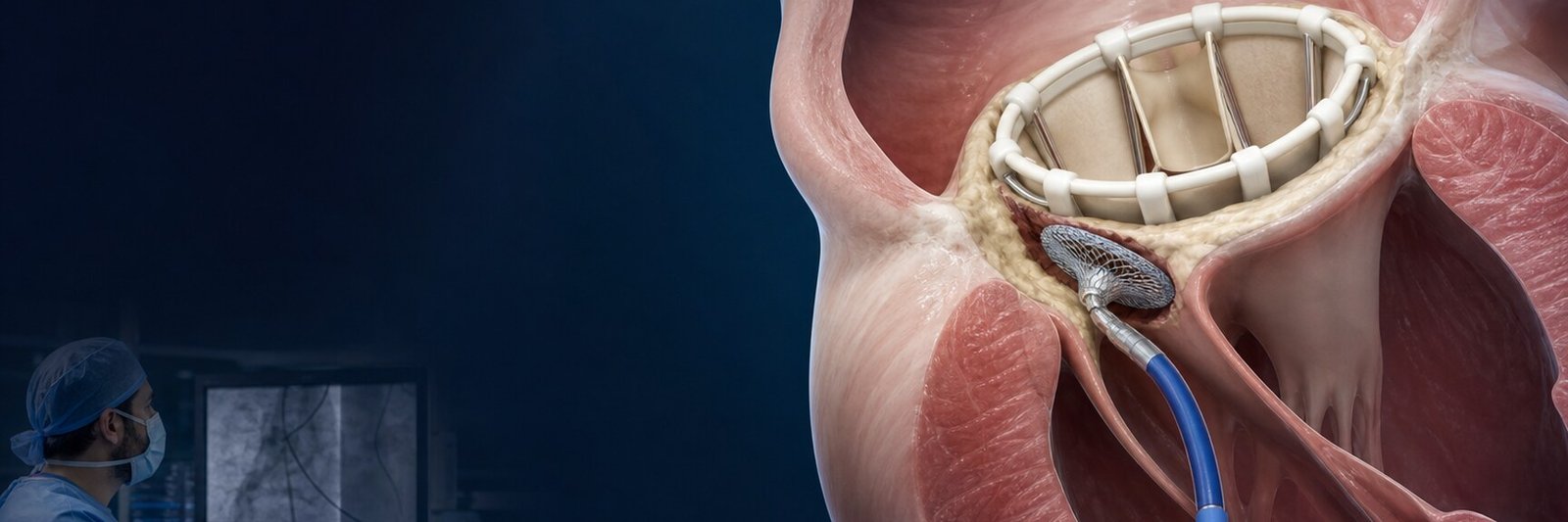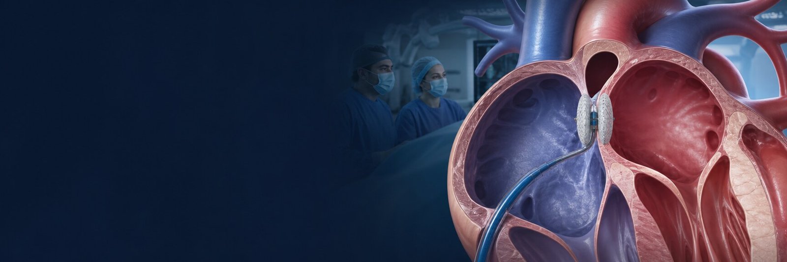
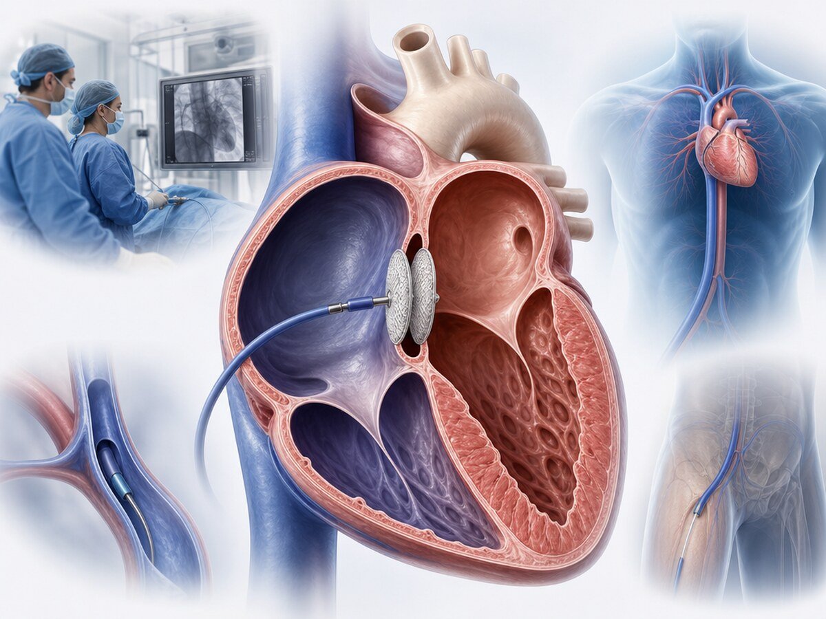
ASD closure is closure of a congenital opening between the heart’s upper chambers. In suitable anatomy a closure device is delivered through a catheter, usually from a femoral vein. The name means atrial septal defect closure. This page is about adults, especially a secundum ASD and transcatheter device closure. Finding an ASD is not, by itself, a reason to close it. The decision follows the size of the shunt, the load on the right heart, pulmonary vascular resistance and left-heart disease.[1,2]
What is an ASD?
An atrial septal defect is a congenital opening in the wall between the right and left atria. Oxygenated blood can pass from the left atrium to the right atrium. That left-to-right shunt, if continuous and large enough, can volume-load the right atrium and right ventricle and increase pulmonary blood flow.[1,4]
A small ASD may stay silent for years. A larger or haemodynamically important opening can cause breathlessness, fatigue, palpitations or arrhythmia. In some adults the finding is incidental on echocardiography done for another reason.[4]
Calling an ASD a “hole in the heart” does not describe one anatomy. Location, rim tissue, pulmonary venous connections and the direction of flow change both the diagnosis and the route of closure.[1]
Are ASD and PFO the same?
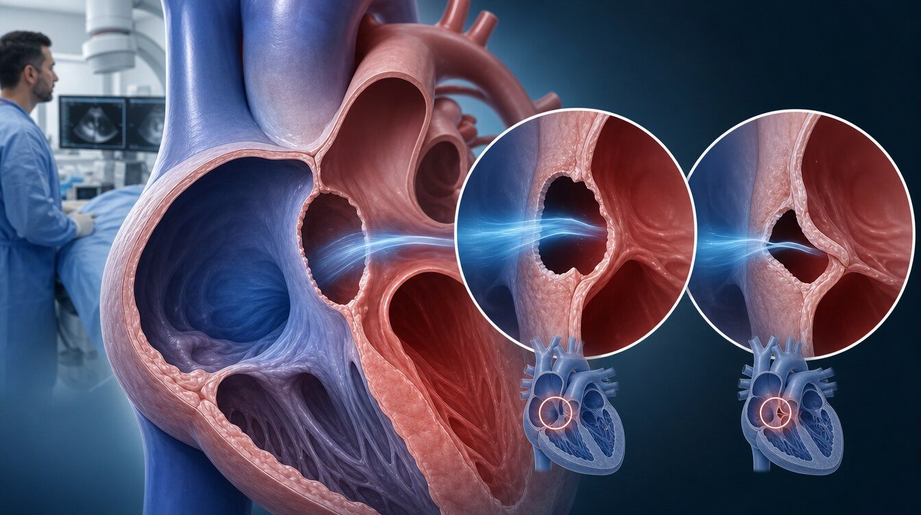
No. An ASD and a PFO (patent foramen ovale) both involve the atrial septum, but they are not the same condition.
In an ASD part of the septum never formed; there is a true gap. Flow is often continuously left to right and the right heart can enlarge.[1,4]
In a PFO the passage needed before birth did not seal completely. Most PFOs are not a continuous wide hole; flow may appear with coughing or straining. A PFO is common; it may remain in about one in four adults and is not closed in most people. The PFO-closure question is usually about selected stroke assessment, which is a different clinical problem from ASD shunt and right-heart load.[4,12]
PFO closure and ASD closure are therefore not interchangeable. Even when a similar device family is used, the question, the anatomy and the follow-up differ.
What are the types of ASD?
In adults the most common type is a secundum ASD, in the fossa ovalis. The 2025 ACC/AHA guideline places about 80 percent of isolated ASDs in this group. Other types sit elsewhere and are not usually suitable for a standard secundum device.[1,14]
- Secundum ASD: an opening in the fossa ovalis. If the rims are adequate and the pulmonary veins connect normally, a transcatheter device may be considered.
- Primum ASD: a defect in the lower septum; it belongs to the partial atrioventricular septal-defect family. Because of the valve relationship it is not treated with a standard secundum device; surgical repair is the usual path.[1,2,14]
- Sinus venosus ASD: near the superior or inferior vena-cava junction, often with anomalous pulmonary venous return. A standard secundum device does not fit. Surgery is the mainstay. In selected superior sinus venosus cases, 2025 ACC/AHA discusses transcatheter closure at expert centres as a conditional, lower-level option; that is not a routine secundum-device procedure.[1,14]
- Coronary-sinus (unroofed coronary sinus) defect: a rare opening of the coronary-sinus roof. It is not suitable for a standard secundum device; surgery or selected expert-centre assessment is required.[1,14]
A catheter-closure plan should not be assumed before the type is clear. Imaging must also review rims and pulmonary venous connections.
What symptoms and later effects can appear?
A small-shunt ASD can stay silent. A significant left-to-right shunt may, over time, lead to:[1,4]
- breathlessness on effort or easy fatigue
- palpitations, atrial fibrillation or another atrial arrhythmia
- right-heart enlargement
- reduced exercise capacity
- rarely, paradoxical embolism when right-to-left passage occurs
An unrepaired significant shunt can, in some people, progress to pulmonary arterial hypertension. In a late stage the shunt can reverse; that is Eisenmenger physiology. That picture is not suitable for closure.[1,3]
Absence of symptoms does not prove that the shunt is unimportant. The decision is read with haemodynamics, right-heart size and pulmonary vascular resistance, not with symptoms alone.[1,2]
Who may be considered?
According to the 2025 ACC/AHA guideline, in adults with an unrepaired ASD, a significant left-to-right shunt (Qp:Qs ≥1.5) and right-ventricular dilation, closure is recommended to improve functional class and outcomes when pulmonary arterial hypertension is absent (PVR ≤2 Wood units) and important left-ventricular disease is absent.[1]
For an isolated secundum ASD with suitable anatomy, a transcatheter approach is usually preferred to surgical repair to shorten stay and recovery. That does not make surgery unsuitable for everyone; another required cardiac operation or inadequate rims can favour surgery.[1,6]
If there is evidence or strong suspicion of paradoxical embolism, 2025 ACC/AHA recommends closure to reduce recurrent embolism when significant PAH or left-heart disease is absent. That frame can apply even if the shunt is not large; it is still not a rule that every stroke leads to ASD closure.[1]
ESC 2020 also recommends closure, even without symptoms, when there is right-ventricular volume overload and no PAH or left-heart disease. A device is the method of choice when a secundum ASD is technically suitable.[2]
A systematic review prepared for the 2018 guideline reported observational improvement in functional capacity and right-heart size; a definite reduction in adult mortality was not shown. That review belongs to the 2018 document, which 2025 ACC/AHA replaces. Benefit is not guaranteed.[1,11]
When is routine closure not appropriate?
Closure is usually not a routine or appropriate option when:[1,2,3]
- the ASD is haemodynamically unimportant and does not load the right heart, and there is no other indication
- Eisenmenger physiology is present: resistance is high enough that the shunt is right to left; closure is considered harmful
- important left-ventricular diastolic disease is judged likely to raise filling pressure dangerously after closure
- the anatomy is primum, most sinus venosus or coronary sinus, and a standard secundum device is proposed
- rims, size or pulmonary venous connections do not fit a device, yet device closure is still insisted upon
“Every ASD is closed” is not a fair summary. The decision is not based on seeing an opening alone, without pulmonary vascular resistance.
How does pulmonary vascular resistance affect the decision?
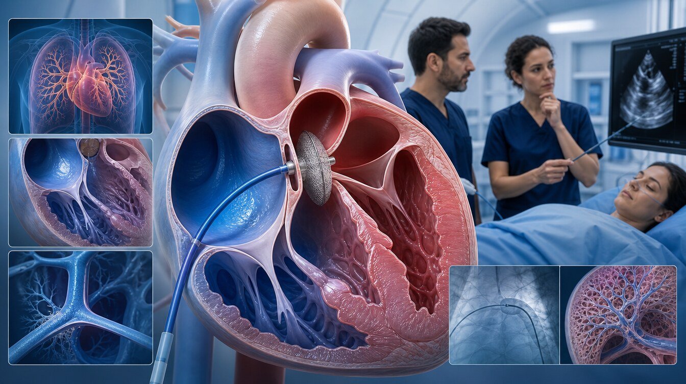
Pulmonary vascular resistance (PVR) is the resistance of the lung vessels, expressed in Wood units (WU). The 2022 ESC/ERS pulmonary-hypertension guideline defines pulmonary arterial hypertension as a mean pulmonary-artery pressure >20 mm Hg, a pulmonary-artery wedge pressure ≤15 mm Hg and PVR >2 Wood units. Every adult with an ASD should be assessed for PAH before closure is planned.[1,3]
For a significant shunt and right-ventricular enlargement, 2025 ACC/AHA reads PVR as follows:[1]
- PVR ≤2 WU (no PAH) and no important left-heart disease: closure is recommended.
- PVR >2 to <5 WU and no important left-heart disease: closure is reasonable to improve functional status.
- PVR 5–8 WU: closure can be beneficial if targeted PAH therapy can bring PVR below 5 WU — a treat-and-reassess path. In the same band, the usefulness of fenestrated repair is more uncertain.
- In Eisenmenger physiology, closure should not be performed.
ESC 2020 uses different cut-offs and wording. Its Class I “no PAH” statement uses invasive confirmation of PVR <3 WU. With PVR 3–5 WU and a significant left-to-right shunt (Qp:Qs >1.5), closure should be considered. With PVR ≥5 WU, targeted PAH therapy comes first; if PVR then falls below 5 WU and a significant shunt remains, fenestrated closure may be considered. Closure is avoided in Eisenmenger physiology or with clear desaturation on exercise.[2]
That difference is not one universal PVR number. 2025 ACC/AHA aligns its “no PAH” threshold with the 2022 PAH definition at 2 WU; ESC 2020 keeps the 3 WU frame of its period. The decision is read with the guideline being used and with that person’s invasive haemodynamics.[1,2,3]
What about left-heart disease and balloon occlusion?
If left-ventricular relaxation (diastolic function) is impaired, especially in older adults, the ASD can act as a pop-off for the left atrium. After closure, left-atrial and left-ventricular filling pressure may rise; breathlessness or pulmonary oedema can worsen.[1,2]
ESC 2020 recommends balloon test occlusion in left-heart disease and a balanced choice among complete closure, fenestrated closure or no closure.[2]
2025 ACC/AHA lists balloon test occlusion before closure as something that may be reasonable to assess haemodynamic change. The supportive text also notes that the test does not always add information beyond a resting left-ventricular end-diastolic pressure and, in some analyses, can overestimate wedge pressure. “The test was done, therefore close” or “the test was not done, therefore do not close” is not a sound rule.[1]
Important left-heart disease in 2025 ACC/AHA includes any condition with chronically elevated left-atrial pressure ≥15 mm Hg. The decision in those people is individual.
How is the assessment planned?
The question is not only “is there a hole?” but type, rims, shunt, right-heart load, rhythm and pulmonary vascular resistance together.[1]
- Transthoracic echocardiography (TTE): first look from the chest wall; right-heart size and estimated pulmonary pressure.
- Transoesophageal echocardiography (TEE): more detail on the whole septum, rims and some pulmonary venous connections.
- Cardiac MR (CMR) or cardiac CT: recommended by 2025 ACC/AHA to define defect size, morphology, rims and pulmonary venous connections. CMR can also quantify the shunt; CT helps with remote venous anomalies.
- Intracardiac echocardiography (ICE): used in some centres during the procedure instead of, or with, TEE; it is not a universal requirement.
- Electrocardiography and rhythm investigation: atrial arrhythmia is common; the pre-closure rhythm plan is individual.
- Cardiac catheterisation: needed to measure PVR when PAH is suspected. ESC 2020 makes invasive PVR mandatory when non-invasive signs suggest raised pulmonary pressure.[1,2]
Inadequate rims, a very large defect or anomalous pulmonary venous return remove a standard secundum device from consideration. Other closures in structural heart interventions — LAA closure and PVL closure — answer different questions.
How is the procedure performed?
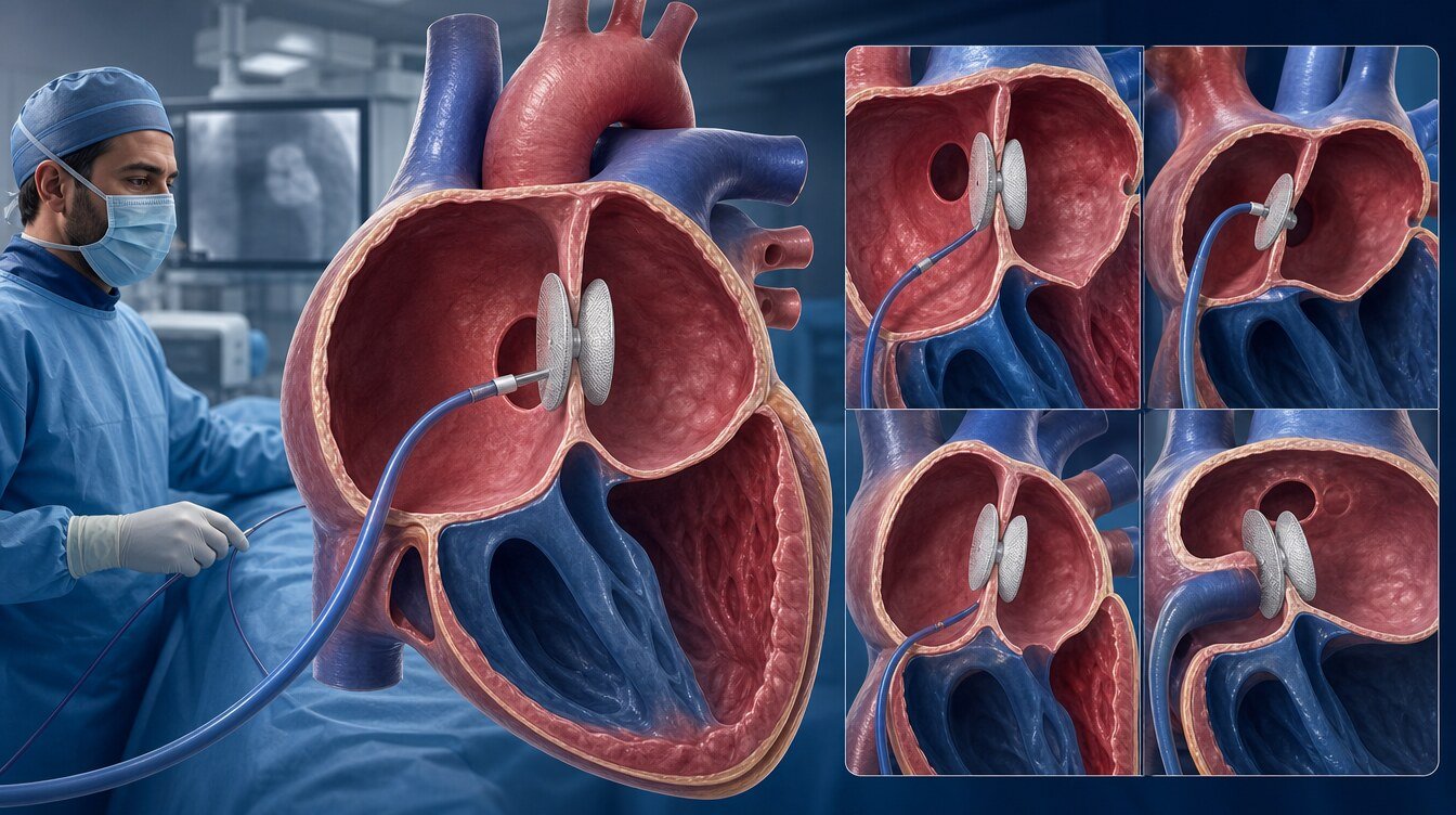
Details vary with anatomy, device and centre protocol. For a secundum ASD the general sequence is:[1,2,6]
- The patient is reviewed again; the sedation or anaesthesia plan is explained.
- Venous access is usually obtained in the groin.
- The catheter is advanced to the right atrium.
- Under imaging, the catheter crosses the defect into the left atrium.
- One part of the device opens on the left side of the septum and the other on the right, grasping both faces.
- Position, rim apposition and residual flow are checked; the device can be repositioned or not released if the result is poor.
- The catheter is removed, pressure is applied at the access site and a period of observation follows.
This is not open-heart surgery. Avoiding a chest incision does not make the procedure simple, risk-free or suitable for everyone. A web page should not be used to derive one universal duration, fasting time or discharge day.
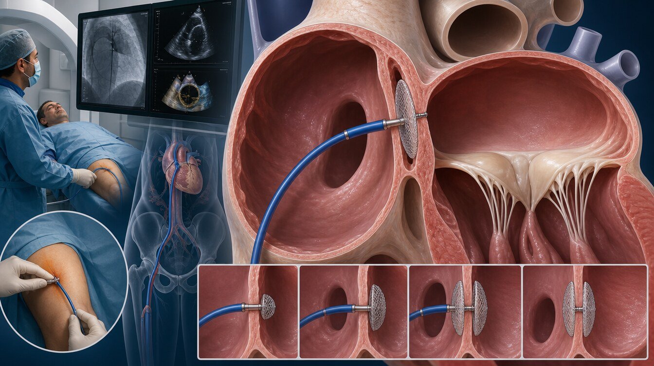
What is an ASD closure device?
An ASD closure device is an implant that opens on both sides of the defect and holds the septal tissue. The aim is to reduce or stop left-to-right flow. Position and apposition are checked with imaging during placement.[1,6]
The device is a permanent implant. Heart tissue is expected to cover it over weeks or months (endothelialisation). Antiplatelet medicines may be used in that period to lower the chance of clot on the device surface. Drug type and duration vary; one prescription should not be copied from a website.[2,10]
Secundum anatomy is not uniform. Device choice depends on rims, defect diameter and intra-procedural checks. This page does not compare brands or give a sizing recipe.
Surgical repair is considered when a device will not fit or another cardiac operation is needed. Comparative studies have reported shorter stay and fewer of some complications with transcatheter closure, while residual shunt and reintervention vary by study. Those results are not a personal success promise.[1,6]
What are the risks?
ASD closure is an invasive procedure whose complication risk is not zero. Individual risk depends on anatomy, device, other illness and how follow-up is done.[1,2,7]
Risks that are discussed include:
- groin bleeding, bruising, pain, venous thrombosis or, rarely, vessel injury
- palpitations, atrial fibrillation or another atrial arrhythmia
- device embolisation or malposition
- clot on the device
- residual shunt after closure
- rarely, device erosion or perforation of the heart or aortic wall
- pericardial effusion
- infection at the access site or, more rarely, related to the device
- contrast allergy or a temporary change in kidney function
ESC 2020 reports that serious complications in suitable secundum cases have been observed at a low rate, and that erosion and thromboembolism appear very rare. A meta-analysis of 28,142 patients reported peri-procedural serious complications in about 1.4 percent; the most frequent serious event was device embolisation, which sometimes needed surgery. Published rates belong to study definitions; they are not a personal calculation.[2,8]
Device erosion
Erosion is thinning or perforation from device pressure on a neighbouring cardiac or aortic wall. It is rare; it should be neither magnified nor dismissed. Case-control data and long-term reviews have linked some anatomical and device features with risk. That is not a rule that every deficient rim will erode or that every adequate rim has zero risk.[7,10]
Residual shunt and rhythm
A small residual passage can be seen on follow-up echocardiography. It does not automatically mean the procedure “failed”; the amount of shunt and the clinical picture are read together.[1,6]
Atrial arrhythmia can be more frequent when an ASD is closed later in life. New atrial fibrillation can appear after the procedure; closure alone does not restore sinus rhythm when a lasting arrhythmia is already present. Closure does not prevent every future rhythm problem.[1,9,15]
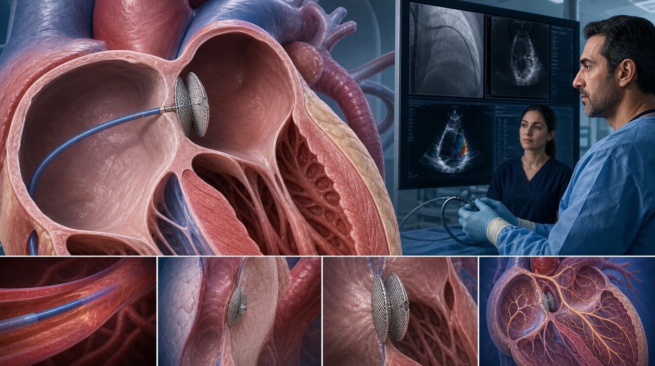
How are recovery, medicines and follow-up arranged?
Observation time, same-day or short hospital stay and return to daily life vary. Mild groin pain or bruising can occur in the first days. Rapidly enlarging swelling, ongoing bleeding, severe pain, fainting, breathlessness, chest pain or marked palpitations need medical review.
Most protocols use antiplatelet therapy until the device is covered by tissue. ESC 2020 describes a period of antiplatelet therapy after device closure; duration and drug choice are not a universal prescription. Anticoagulants are not started routinely for an ASD alone; they may be needed if atrial fibrillation or another reason is present.[2]
Antibiotic protection against endocarditis may be discussed in the early months after prosthetic material, or longer if a residual shunt remains. That is not one universal dental recipe; the current endocarditis guideline and the person’s residue are read together.[2]
Follow-up checks device position, residual shunt, right-heart size, pulmonary pressure and rhythm. ESC 2020 considers regular review for the first two years after a device, then longer intervals depending on findings, and regular ACHD follow-up when closure is done in adulthood, especially after age 40. 2025 ACC/AHA steps intervals by physiological stage. The calendar should not be copied from this page.[1,2]
Sudden facial droop, arm or leg weakness, speech difficulty or sudden visual loss may be stroke symptoms. In Türkiye, call 112.
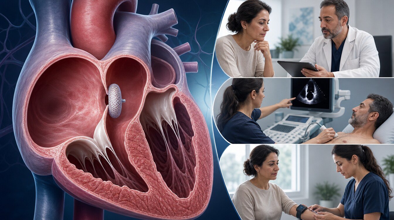
Pregnancy and older age
A haemodynamically important unrepaired ASD can increase volume load in pregnancy. A small ASD that does not load the heart is often well tolerated. Series of repaired ASD often report low pregnancy complication rates; that is not a guarantee that every pregnancy is low risk. Severe pulmonary hypertension or Eisenmenger physiology is considered high risk. This page does not write a pregnancy ban or a universal “close then conceive” rule. If closure is considered, timing, pregnancy plans and PAH are discussed together.[1,2,13]
Older age does not automatically open or close the door to the procedure. A randomised trial showed that surgical closure after age 40 could reduce some clinical events compared with medical observation; that is not a mandate to treat every older adult. Other illness, rhythm, PVR and left-heart filling pressure remain part of the decision, independent of age alone.[1,5]
What happens if the ASD is not closed?
A small ASD that does not load the right heart may cause no problem if left open.[4]
If a significant left-to-right shunt continues, right-heart volume load, reduced exercise capacity, atrial arrhythmia and, in some people, pulmonary vascular disease remain possible. Not closing does not mean Eisenmenger physiology will certainly develop. Closing also does not remove every future rhythm problem or every cause of stroke.[1,2]
The choice is not only “close the hole” versus “do nothing”; it is individualised among complete closure, fenestrated closure, PAH therapy and observation.
Frequently asked questions
The usual frame is a Qp:Qs of 1.5 or more, right-ventricular enlargement, no PAH or an acceptable PVR, and no important left-heart disease. Transcatheter closure is usually preferred for a suitable isolated secundum ASD.[1]
Antiplatelet type and duration depend on the device, the protocol and bleeding risk. One prescription should not be copied from a web page.[2]
Assessment and decision
The ASD closure decision does not rest on the sentence “there is a hole in the heart.” Defect type, rims and pulmonary veins, Qp:Qs and right-ventricular size, the PVR band, left-heart filling pressure, rhythm and the person’s preferences are read together. ESC 2020 and 2025 ACC/AHA point in the same direction but do not write the same PVR number.[1,2,3]
Literature
References
Gurvitz M, et al. 2025 ACC/AHA/HRS/ISACHD/SCAI Guideline for the Management of Adults With Congenital Heart Disease. Circulation. 2025. doi:DOI: 10.1161/CIR.0000000000001402. The same document in JACC: doi:DOI: 10.1016/j.jacc.2025.09.006 (not a separate body of evidence). It replaces the 2018 AHA/ACC ACHD guideline.
Baumgartner H, et al. 2020 ESC Guidelines for the management of adult congenital heart disease. European Heart Journal. 2021. doi:DOI: 10.1093/eurheartj/ehaa554. PMID 32860028.
Humbert M, et al. 2022 ESC/ERS Guidelines for the diagnosis and treatment of pulmonary hypertension. European Respiratory Journal. 2022. doi:DOI: 10.1183/13993003.00879-2022.
American Heart Association. Atrial Septal Defect (ASD). Official patient information. heart.org.
Attie F, et al. Surgical treatment for secundum atrial septal defects in patients >40 years old. A randomized clinical trial. J Am Coll Cardiol. 2001. doi:DOI: 10.1016/S0735-1097(01)01635-701635-7).
Du ZD, et al. Comparison between transcatheter and surgical closure of secundum atrial septal defect in children and adults. J Am Coll Cardiol. 2002. doi:DOI: 10.1016/S0735-1097(02)01862-401862-4). PMID 12039500.
McElhinney DB, et al. Relative Risk Factors for Cardiac Erosion Following Transcatheter Closure of Atrial Septal Defects. Circulation. 2016. doi:DOI: 10.1161/CIRCULATIONAHA.115.019987.
Abaci A, et al. Short and long term complications of device closure of atrial septal defect and patent foramen ovale: Meta-analysis of 28,142 patients from 203 studies. Catheterization and Cardiovascular Interventions. 2013. doi:DOI: 10.1002/ccd.24875.
Himelfarb J, et al. Atrial fibrillation following transcatheter atrial septal defect closure: a systematic review and meta-analysis. Heart. 2021. doi:DOI: 10.1136/heartjnl-2021-319794.
Jalal Z, et al. Long-term Complications After Transcatheter Atrial Septal Defect Closure: A Review of the Medical Literature. Canadian Journal of Cardiology. 2016. doi:DOI: 10.1016/j.cjca.2016.02.068.
Oster M, et al. Interventional Therapy Versus Medical Therapy for Secundum Atrial Septal Defect: A Systematic Review (Part 2) for the 2018 AHA/ACC Guideline. J Am Coll Cardiol. 2018. doi:DOI: 10.1016/j.jacc.2018.08.1032. The Circulation version of the same review is not a separate body of evidence.
Kheiwa A, et al. Patent foramen ovale and atrial septal defect. Echocardiography. 2020. doi:DOI: 10.1111/echo.14646.
Bredy C, et al. Pregnancy in adults with repaired/unrepaired atrial septal defect. Journal of Thoracic Disease. 2018. doi:DOI: 10.21037/jtd.2017.10.130.
Fraisse A, et al. Atrial septal defect closure: indications and contra-indications. Journal of Thoracic Disease. 2018. doi:DOI: 10.21037/jtd.2018.08.111.
Duong P, et al. Atrial arrhythmia after transcatheter closure of secundum atrial septal defects in patients ≥40 years of age. Europace. 2016. doi:DOI: 10.1093/europace/euw186.
This content is for general information only and does not replace personalized medical advice. In emergencies call local emergency services.
Continue