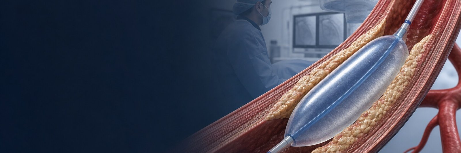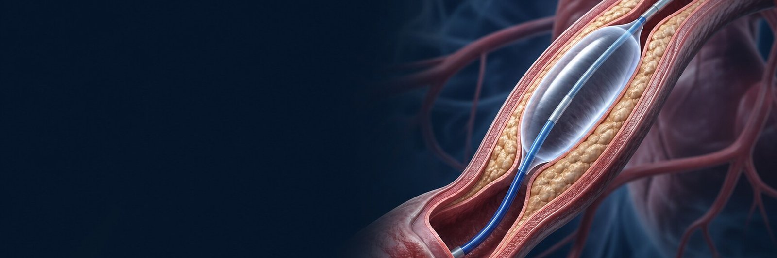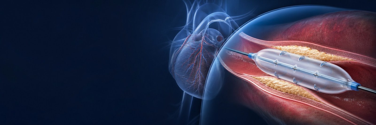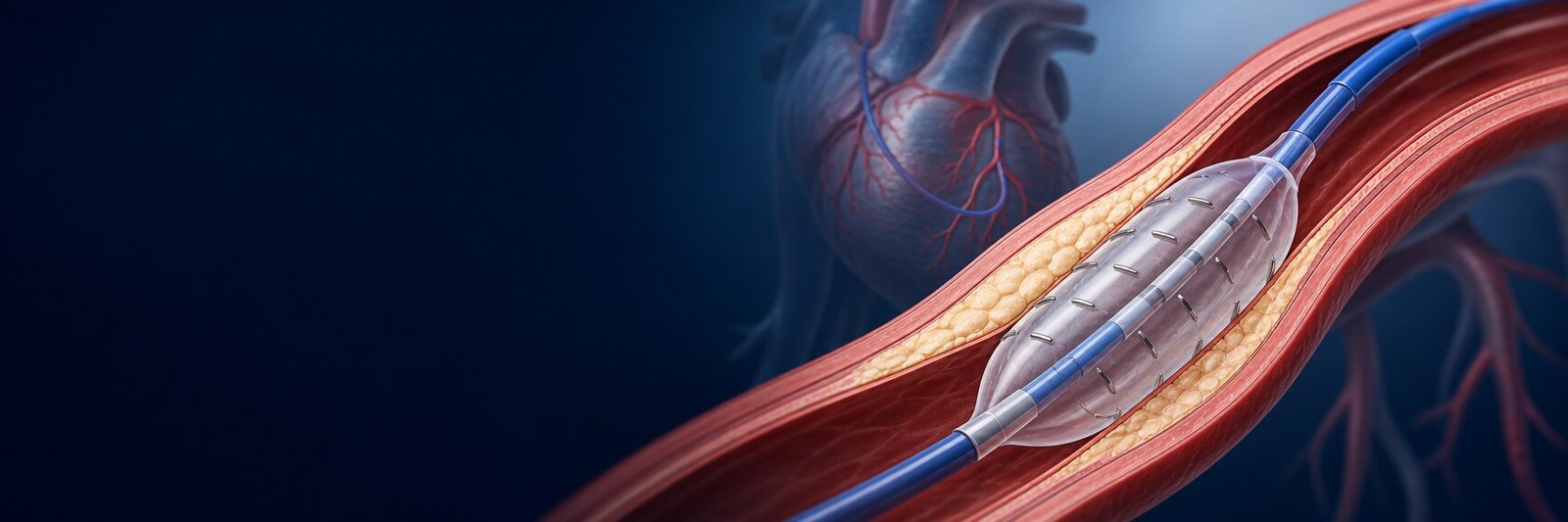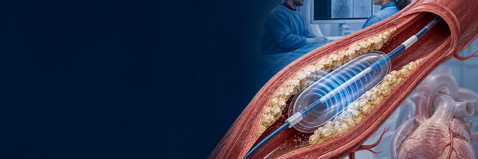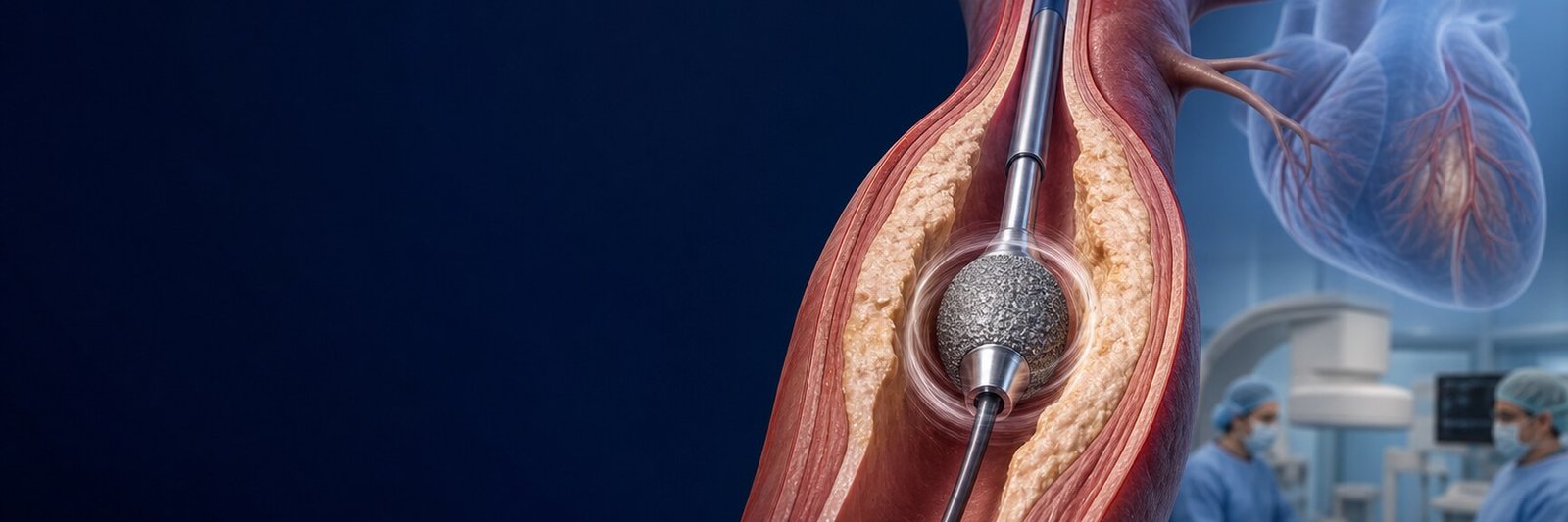
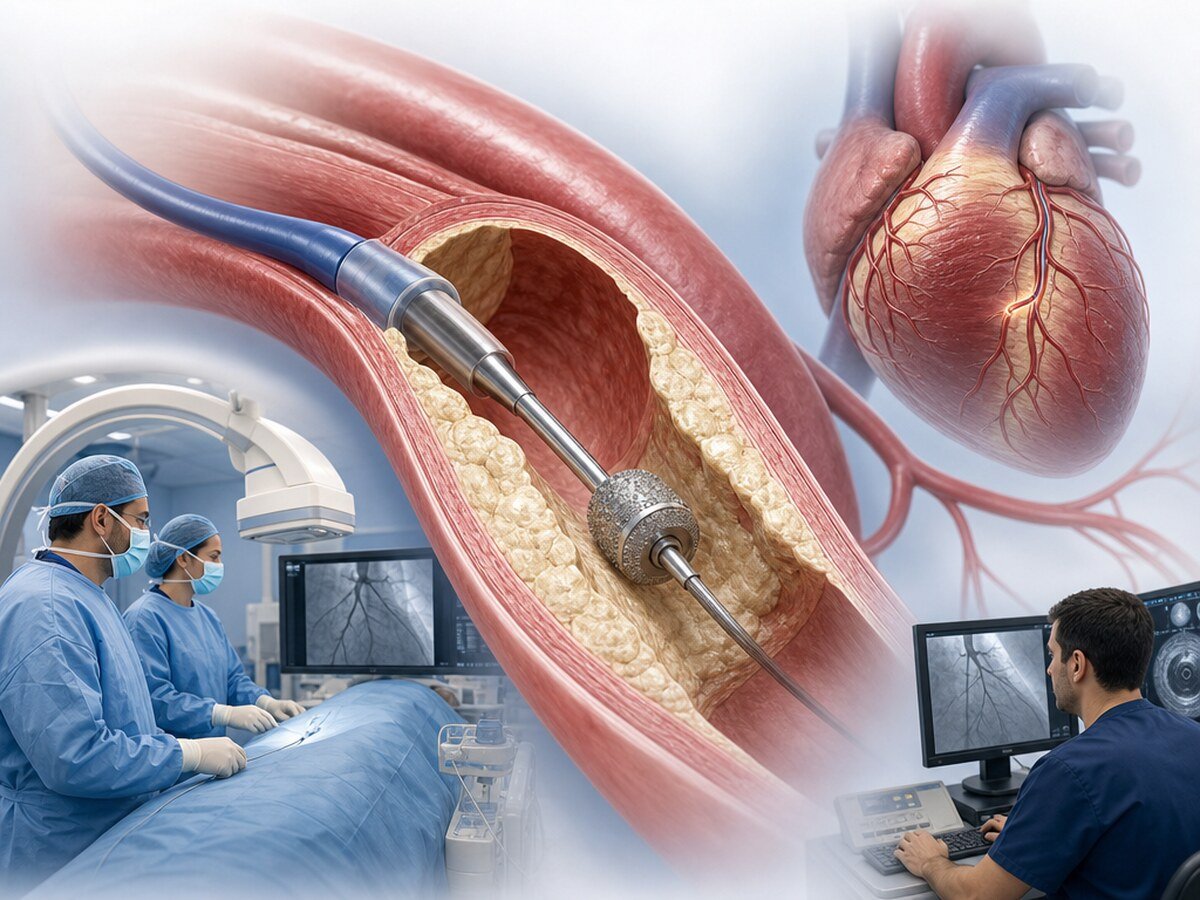
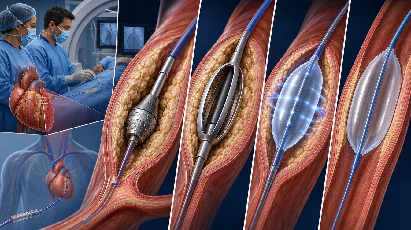
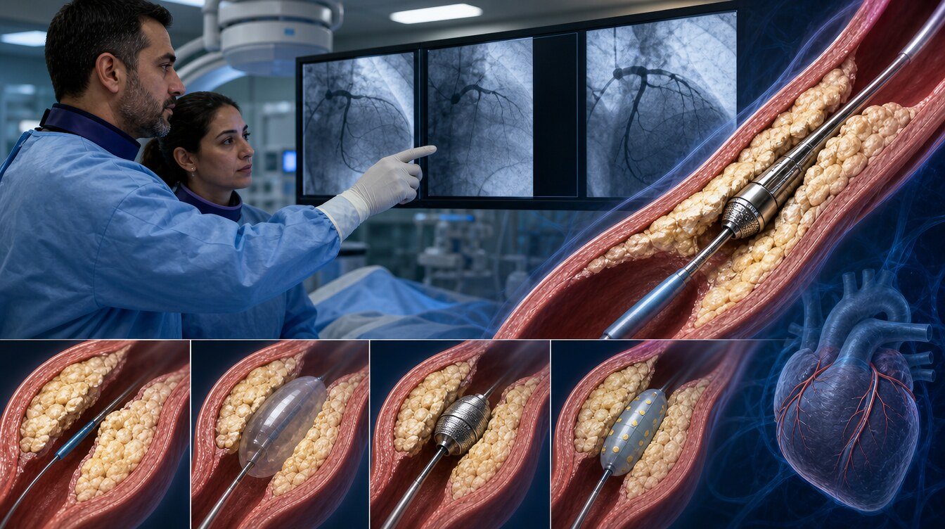
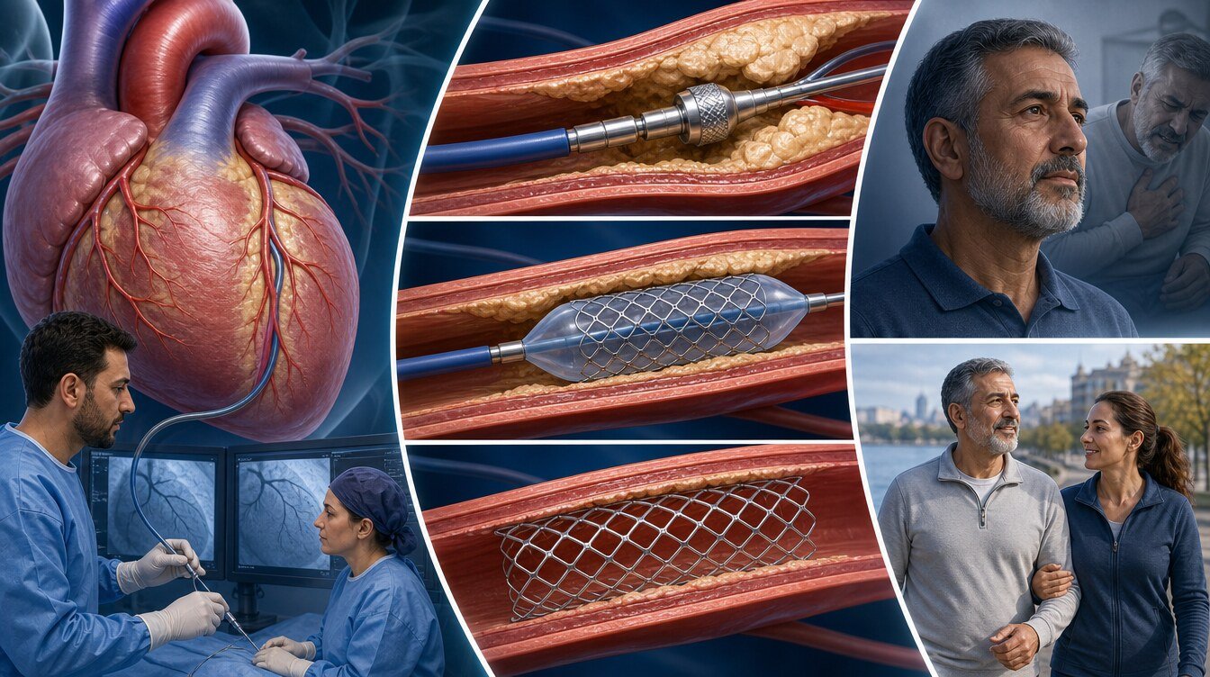
Rotational atherectomy (rotablation) is a temporary intravascular tool used to prepare a hard, calcified coronary stenosis for a stent or another treatment. A diamond-coated burr advanced over a fine guidewire rotates in a controlled way to abrade plaque and help open a path in the lumen. It is removed afterwards and is not left in the artery. It is not required in every calcified stenosis, and plaque is not completely cleared.[14,16,18]
What does rotational atherectomy mean?
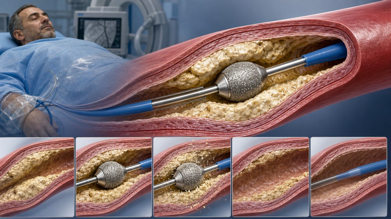
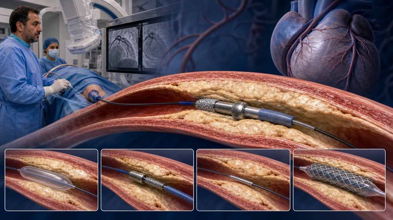
Rotational atherectomy aims to abrade hard tissue with a rotating diamond-coated tip (burr). The goal is to prepare a path so that a balloon or stent can be delivered and expanded more controllably in a selected stenosis. That does not mean complete removal of calcium, drilling a tunnel or a guaranteed fully expanded stent.[16,18]
Burr diameter, rotational speed and duration are device- and lesion-specific. This page therefore does not publish a universal burr size, speed, duration or pressure recipe. Rotational atherectomy is not a permanent implant like a stent; after preparation the burr is withdrawn with the catheter.[18]
Why is it sometimes called a “drill”?
In everyday language, “drill” is a misunderstanding of the rotating tip. The device does not bore a tunnel like a construction drill and does not destroy calcium. It performs controlled plaque modification and path preparation with a diamond-coated burr. It is not a category name and not a frightening marketing phrase.[14,18]
When may it be considered?
Rotational atherectomy is not a routine step in every calcified stenosis. Use depends on whether a balloon or imaging catheter can cross the lesion, calcium depth and circumference, the stent plan, and operator and centre experience.[14,16,17]
Calcified stenoses that are hard to cross or to open
In selected severely calcified lesions where a balloon cannot cross, or crosses but cannot expand adequately, rotational atherectomy may be considered to help stent delivery and expansion. That does not mean it is required in every calcified artery.[14,16]
Calcium phenotype selected with imaging
IVUS or OCT can help distinguish whether calcium is superficial or deep and how far it encircles the vessel. Phenotype may influence the choice among rotational atherectomy, balloons, IVL or a combined strategy; an image alone does not mandate the method.[16,17]
Tissue inside a prior stent (historical, a different question)
In the older POBA/bare-metal-stent era, rotational atherectomy was also studied for tissue inside a stent (ISR). That is not the same question as a new (de novo) calcified stenosis in the contemporary drug-eluting-stent era, and the results conflict.[9,10]
When may it be unsuitable or insufficient alone?
Rotational atherectomy does not produce an adequate result in every lesion. It should not be presented as a routine method in thrombotic, freshly occlusive (for example selected STEMI) lesions; the decision is individual.[16] It may not be enough alone in deep circumferential calcium, calcium beneath an under-expanded stent, or when another mechanism dominates.[16,17]
A different strategy may be needed when:
- The device cannot cross the stenosis safely
- The burr advances but preparation remains inadequate
- Slow flow or no-reflow develops
- Vessel-wall injury, perforation or flow-limiting dissection occurs
- Resistant calcium beneath a stent needs another method
- Operator or centre experience is not suited to this tool
A standard NC, scoring or cutting balloon, a super high-pressure balloon, IVL, laser or a combined approach may then be considered. These tools are not universal substitutes for one another.[8,16,17]
Why may IVUS or OCT be used?
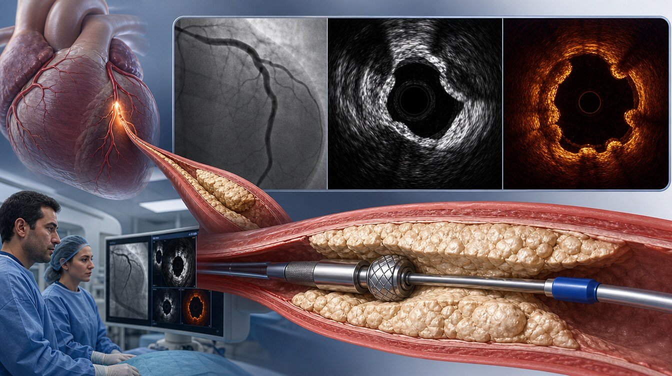
Coronary angiography gives a two-dimensional lumen view and can miss calcium depth, circumferential extent and true stent expansion. Intravascular ultrasound (IVUS) and optical coherence tomography (OCT) provide cross-sectional images that can help assess:[16,17]
- Whether calcium is superficial or deep, and how far it encircles the vessel
- Whether the burr, balloon or stent has created an adequate path or expansion
- Whether the stent is apposed to the wall
- Whether restenosis is tissue inside the stent or calcium beneath it
Consensus texts discuss imaging as a guide to calcium phenotype and preparation choice. That does not mean imaging prevents heart attack or death, that it mandates rotational atherectomy, or that it is technically possible in every patient. No universal burr or expansion cut-off is published here.[16,17]
How is the procedure performed?
The procedure is done in a catheter laboratory equipped for coronary angiography and percutaneous coronary intervention (PCI). It is not open-heart surgery; the patient experience is similar to other coronary balloon–stent procedures:[19]
- Preparation: History, medicines, blood tests, kidney function and contrast risk are reviewed.
- Access: Radial or femoral artery under local anaesthesia.
- Imaging and wire: The artery is imaged; a fine wire is advanced beyond the stenosis.
- Preparation: The team chooses rotational atherectomy, a balloon, IVL, a stent or another tool according to the lesion and the live result. The burr is removed at the end.
- Result check: Flow, residual stenosis and, when used, IVUS/OCT findings are reassessed.
- Removal: Wires and catheters are withdrawn; the access site is controlled. If a stent was placed, the lasting structure is the stent.
Most patients remain awake. Brief chest discomfort during the procedure should be reported if new or severe.[19]
This page does not prescribe burr diameter, rotational speed, duration or device order.
What does the scientific evidence show?
Three ideas should be kept apart. Strategy or procedural success is whether the planned approach—including a bailout tool if needed—treats the lesion adequately. Stent delivery and acute lumen gain are mechanical results at the time of the procedure. Late lumen loss, death, myocardial infarction and repeat procedures are separate, more patient-important endpoints. They are not interchangeable.[1,2]
ROTAXUS: strategy success versus angiographic and clinical results
ROTAXUS randomised 240 patients to planned rotational atherectomy plus a paclitaxel-eluting stent or to a stenting strategy without planned rotational atherectomy; crossover was allowed (120/120). Strategy success was higher with planned rotational atherectomy (92.5% versus 83.3%). Acute lumen gain was 1.56 mm versus 1.44 mm. The primary endpoint—nine-month in-stent late lumen loss—was not reduced by rotational atherectomy. Target-lesion revascularisation (11.7% versus 12.5%) and definite stent thrombosis (0.8% versus 0%) were similar. The trial was not powered for death or myocardial infarction. A separate stent-delivery percentage was not verified in this package. It is a paclitaxel-stent-era trial and is not evidence of routine clinical superiority.[1]
The two-year ROTAXUS paper is follow-up of the same 240-person cohort. A DOI/PMID was not verified in this package and numerical results were not re-extracted, so it is not used as an independent trial or as extra proof of superiority.
The PREPARE-CALC family: mixed modified balloon, the same 200 people
PREPARE-CALC compared rotational atherectomy with a mixed modified-balloon strategy in 200 severely calcified native lesions (100/100). The modified arm combined scoring and cutting balloons; bailout rotational atherectomy was allowed. It is not a single-balloon-device trial. Strategy success was 98% with rotational atherectomy versus 81% with modified balloons. At nine months, target-lesion revascularisation was 2% versus 7% (not statistically significant); stent thrombosis was 0% in both arms. The trial was not powered for superiority in death or myocardial infarction.[2]
The two-year paper is follow-up of the same 200-person family (97% follow-up). Death (8% versus 7%), myocardial infarction (5% versus 4%) and target-vessel revascularisation (9% versus 5%, modified versus rotational atherectomy) did not differ significantly. The authors note that the sample is too small for definitive clinical-endpoint conclusions.[3]
The five-year paper is again the same 200-person cohort, not independent replication. Target-vessel failure was similar (21% rotational atherectomy, 19% modified balloon); target-lesion revascularisation was lower after rotational atherectomy (3% versus 12%). That is a long-term signal from the same small family.[4]
PREPARE-CALC OCT is a 122-person imaging subset of the same family. Preparation strategy was reported not to determine OCT stent expansion; it is not a separate 122-person clinical trial.[5]
ROTA.shock: rotational atherectomy versus IVL, an imaging endpoint
The ROTA.shock parent study randomised 70 patients 1:1 to rotational atherectomy or coronary IVL. The primary endpoint is minimum stent area (imaging). MSA was 6.60 mm² (RA) versus 6.10 mm² (IVL); IVL was noninferior for MSA and stent expansion was similar. The trial was not powered for death or myocardial infarction. Lesion crossability and crossover were not extracted as separately verified endpoints in this package; OCT area is not the same as the ability to cross the stenosis. A single trial does not establish universal superiority.[7]
The 21-person OCT plaque-modification paper is a nested report of the same 70-person cohort. It describes more/longer calcium fractures with IVL and greater acute lumen gain with rotational atherectomy; it must not be counted as a separate clinical trial.[6]
ROLLER COASTR-EPIC22: three-arm OCT expansion
ROLLER assigned 171 patients to rotational atherectomy, IVL or excimer laser (ELCA) (57 per arm). The primary endpoint is OCT percentage stent expansion. Minimum stent area was 5.5 mm² with rotational atherectomy, 5.4 mm² with IVL and 5.1 mm² with ELCA; IVL was noninferior to rotational atherectomy, while ELCA did not meet the prespecified margin. In the rotational-atherectomy arm there were 2/57 perforations, 1/57 slow/no-reflow, 1/57 dissection (not flow-limiting), 0/57 in-hospital stent thromboses and 0 procedure/in-hospital deaths. These small-arm counts are not a personal risk forecast. The trial carries a crossover and combination-technique limitation; lesion crossability as a separate endpoint is NA in this package.[8]
ROTA-CUT: rotational atherectomy in both arms
ROTA-CUT compared rotational atherectomy followed by a cutting balloon with rotational atherectomy followed by an NC balloon in 60 patients. IVUS minimum stent area was similar (6.7 versus 6.9 mm²). Adding a cutting balloon after rotational atherectomy did not increase stent area versus an NC balloon. This does not answer whether rotational atherectomy is more effective than balloons.[12]
ROSTER and ARTIST: older ISR era
ROSTER (200 patients) and ARTIST (298 patients) studied tissue inside stents in the older POBA/bare-metal-stent era. In ROSTER, nine-month target-lesion revascularisation was lower with rotational atherectomy (32% versus 45%). In ARTIST, six-month restenosis was higher with the rotational-atherectomy strategy (65% versus 51%) and in-hospital myocardial infarction was 4.6% versus 1.4%. Conflicting historical ISR evidence must not be generalised to contemporary drug-eluting-stent de novo calcified stenoses or to calcium outside an under-expanded stent.[9,10]
Registries and CORECT: descriptive or device-versus-device comparison
The J2T registry reports 1,090 patients treated with rotational atherectomy in 2004–2015. Procedural success was 96.2%, in-hospital death 3.0% and definite/probable stent thrombosis 1.3%. There is no comparator arm; the mix includes high-risk cases such as dialysis. These figures are not a patient’s expected risk.[11]
CORECT compared a novel rotational-atherectomy system with an established rotational-atherectomy system in a 15-person first-in-human phase and a 224-person pivotal phase (111 versus 113). IVUS minimum stent area was 5.29 mm² in both arms. This is not evidence that rotational atherectomy is superior to IVL, laser or balloons; it is a device-versus-device noninferiority study. Online/issue display status was not independently verified in the package.[13]
What are possible benefits and limitations?
| Possible benefit / purpose | Limitation / required caveat |
|---|---|
| Path preparation and controlled plaque modification in a selected calcified stenosis | Does not clear plaque completely; does not bore a tunnel; not required in every lesion |
| Higher strategy success in ROTAXUS | Nine-month late lumen loss was not reduced; clinical superiority was not shown |
| Higher strategy success in PREPARE-CALC | Comparator was a mixed modified balloon; no death/MI/TVR difference at 2 years; 5-year data are the same cohort |
| Similar imaging results versus IVL in ROTA.shock and ROLLER | Imaging endpoints; not a universal “which is better” ranking |
| The burr is removed at the end | Most de novo plans still consider a stent |
What are the risks?
Rotational atherectomy is used within coronary intervention. In addition to the general risks of balloon–stent procedures, method-specific complications are described:[8,18,19]
- Coronary dissection
- Coronary perforation
- Slow flow or no-reflow
- Burr entrapment
- Acute vessel closure
- Side-branch loss
- Bradycardia, conduction disturbance or need for temporary pacing
- Myocardial infarction, stent thrombosis or arrhythmia
- Access-site bleeding and vascular complications
- Contrast-related kidney injury or allergy
- Later restenosis if a stent is used
The two perforations and one slow/no-reflow event in the ROLLER rotational-atherectomy arm are small-arm observations and must not be copied as a personal risk calculator.[8] The J2T in-hospital death rate also belongs to a registry population.[11]
What are the alternatives and complementary methods?
| Method | Core approach | Key difference from rotational atherectomy |
|---|---|---|
| Standard NC balloon | Controlled expansion without surface elements | Does not abrade or debulk |
| Scoring / cutting balloon | Focused preparation via surface elements or microblades | Compared as a mixed arm in PREPARE-CALC; not single-device evidence |
| Super high-pressure NC | Selected resistant lesions not opened by a standard NC balloon | A separate balloon class |
| Intravascular lithotripsy (IVL) | Acoustic pressure waves that fracture calcium | The balloon must still cross; imaging equivalence is not universal superiority |
| Orbital atherectomy | A different atherectomy class | Must not be transferred to coronary rotational-atherectomy evidence |
| Excimer laser (ELCA) | Laser ablation | A separate arm in ROLLER; not equated with rotational atherectomy |
| Peripheral atherectomy | Leg/peripheral vascular bed | Outside coronary rotational-atherectomy evidence |
Methods may be combined in the same procedure; combined use is not a routine prescription for every patient. ROTA-CUT showed that adding a cutting balloon after rotational atherectomy did not increase stent area versus an NC balloon.[12,16,17]
Preparation, discharge and recovery
Preparation depends on elective versus urgent setting, kidney function, bleeding risk, medicines, contrast history and access route. Follow hospital fasting and medicine instructions; do not stop prescribed medicines on your own.[19]
After planned uncomplicated coronary angioplasty some patients go home the same or next day. Local policy, clinical status and complications change the timing. This general frame comes from NHS patient information and is not a fixed discharge rule for every hospital in Türkiye.[19]
Discharge advice should be personalised and include access-site care, activity limits, antiplatelet and other prescriptions, follow-up, and when to seek emergency care.
Antiplatelet duration is set by the overall PCI, stent or drug-coated balloon strategy and the clinical picture, not by rotational atherectomy alone. Stopping these medicines without advice can cause serious stent thrombosis.[14,19]
Bruising at the access site can occur. Increasing pain, swelling, redness, discharge or fever should prompt contact with the treating centre or another healthcare service.[19]
When should emergency services be called?
Call emergency services (112 in Türkiye) without delay for:[19]
- Chest pain that does not settle or is worsening
- Access-site bleeding that continues despite direct pressure or starts again
- Marked coldness, colour change or numbness in the treated arm or leg
- Fainting, severe breathlessness, sudden weakness or a change in consciousness
Not every bruise or mild tenderness is an emergency; uncontrolled bleeding, severe chest pain or impaired limb circulation is. Do not drive yourself with emergency symptoms. The 112 direction is a general safety frame; detailed local discharge wording should be reviewed against an official Turkish source before publication.[19]
Frequently asked questions
No. Strategy success was higher with planned rotational atherectomy; nine-month late lumen loss was not reduced and the trial was not powered for death or myocardial infarction. It is not evidence of routine clinical superiority.[1]
No. ROTA.shock is a small comparison with an imaging primary endpoint; IVL was noninferior for minimum stent area. Lesion crossability was not separately verified. A single trial does not establish universal superiority.[7]
Assessment and decision
Rotational atherectomy is a temporary preparation tool used in selected calcified coronary stenoses. Planning is broader than “will rotational atherectomy be used?” and includes treatment need, device crossability, calcium morphology, the stent plan, IVUS/OCT findings, alternatives and centre experience.[14–18]
The 2021 ACC/AHA/SCAI and 2024 ESC documents have no rotational-atherectomy-specific Class/LOE recommendation re-verified in this package (NA). EAPCI, SCAI and Japanese CVIT texts are consensus statements and must not be presented as graded guideline recommendations. The decision rests on individual clinical assessment.[14–18]
Literature
References
Abdel-Wahab M, et al. High-speed rotational atherectomy before paclitaxel-eluting stent implantation in complex calcified coronary lesions: the randomized ROTAXUS trial. JACC Cardiovasc Interv. 2013. DOI: 10.1016/j.jcin.2012.07.017. PMID: 23266232.
Abdel-Wahab M, et al. High-Speed Rotational Atherectomy Versus Modified Balloons Prior to Drug-Eluting Stent Implantation in Severely Calcified Coronary Lesions: The Randomized PREPARE-CALC Trial. Circ Cardiovasc Interv. 2018. DOI: 10.1161/CIRCINTERVENTIONS.118.007415. Trial: NCT02502851. A PMID was not verified in this package.
Allali A, et al. High-speed rotational atherectomy versus modified balloons for plaque preparation of severely calcified coronary lesions: two-year outcomes of the randomised PREPARE-CALC trial. EuroIntervention. 2023. DOI: 10.4244/EIJ-D-22-00677. PMID: 36579635. PMCID: PMC10068860. Trial: NCT02502851.
Mankerious N, et al. Lower revascularization rates after high-speed rotational atherectomy compared to modified balloons in calcified coronary lesions: 5-year outcomes of the randomized PREPARE-CALC trial. Clin Res Cardiol. 2024. DOI: 10.1007/s00392-024-02434-1. PMID: 38483633. Trial: NCT02502851.
Hemetsberger R, et al. Optical Coherence Tomography Assessment in Patients Treated With Rotational Atherectomy Versus Modified Balloons: PREPARE-CALC OCT. Circ Cardiovasc Interv. 2021. DOI: 10.1161/CIRCINTERVENTIONS.120.009819. Trial: NCT02502851. A PMID was not verified in this package. This is an imaging subset of the same 200-person family.
Blachutzik F, et al. Comparison of Coronary Intravascular Lithotripsy and Rotational Atherectomy in the Modification of Severely Calcified Stenoses. Am J Cardiol. 2023. DOI: 10.1016/j.amjcard.2023.02.028. PMID: 37012181. Nested OCT report of the ROTA.shock cohort.
Blachutzik F, et al. Coronary intravascular lithotripsy and rotational atherectomy for severely calcified stenosis: Results from the ROTA.shock trial. Catheter Cardiovasc Interv. 2023. DOI: 10.1002/ccd.30815. PMID: 37668088.
Jurado-Román A, et al. Rotational Atherectomy, Lithotripsy, or Laser for Calcified Coronary Stenosis: The ROLLER COASTR-EPIC22 Trial. JACC Cardiovasc Interv. 2025. DOI: 10.1016/j.jcin.2024.11.012. PMID: 39918495. Trial: NCT04181268.
Sharma SK, et al. Randomized trial of Rotational Atherectomy Versus Balloon Angioplasty for Diffuse In-Stent Restenosis (ROSTER). Am Heart J. 2004. DOI: 10.1016/j.ahj.2003.07.002. PMID: 14691413.
vom Dahl J, et al. Rotational Atherectomy Does Not Reduce Recurrent In-Stent Restenosis: Results of the Angioplasty versus Rotational Atherectomy for Treatment of Diffuse In-Stent Restenosis Trial (ARTIST). Circulation. 2002. DOI: 10.1161/hc0502.103347. PMID: 11827923.
Okai I, et al. Clinical Characteristics and Long-Term Outcomes of Rotational Atherectomy: J2T Multicenter Registry. Circ J. 2018. PMID: 28931790. A DOI was not verified in this package (NA).
Sharma SK, et al. Rotational atherectomy combined with cutting balloon to optimise stent expansion in calcified lesions: the ROTA-CUT randomised trial. EuroIntervention. 2024. DOI: 10.4244/EIJ-D-23-00811. PMCID: PMC10756220. A PMID was not verified in this package.
Li C, et al. Safety and Efficacy of a Novel Rotational Atherectomy System in Coronary Calcifications (CORECT). JACC Asia. 2026. DOI: 10.1016/j.jacasi.2026.04.035. PMID: 42412697. Trial: NCT05447585. Online/issue display status was not independently verified.
Lawton JS, et al. 2021 ACC/AHA/SCAI Guideline for Coronary Artery Revascularization. Circulation. 2022. DOI: 10.1161/CIR.0000000000001038. PMID: 34882435. No rotational-atherectomy-specific Class/LOE was re-verified in this package (NA).
Vrints C, et al. 2024 ESC Guidelines for the management of chronic coronary syndromes. Eur Heart J. 2024. DOI: 10.1093/eurheartj/ehae177. PMID: 39210710. No rotational-atherectomy-specific Class/LOE was directly verified in this package (NA).
Barbato E, et al. Management strategies for heavily calcified coronary stenoses: an EAPCI clinical consensus statement in collaboration with the EURO4C-PCR group. Eur Heart J. 2023. DOI: 10.1093/eurheartj/ehad342. PMID: 37208199.
Riley RF, et al. SCAI Expert Consensus Statement on the Management of Calcified Coronary Lesions. JSCAI. 2024. DOI: 10.1016/j.jscai.2023.101259. PMID: 39132214. PMCID: PMC11307856.
Sakakura K, et al. Clinical expert consensus document on rotational atherectomy from the Japanese association of cardiovascular intervention and therapeutics: update 2023. Cardiovasc Interv Ther. 2023. DOI: 10.1007/s12928-022-00906-7. PMID: 36529831.
NHS. Coronary angioplasty and stent insertion: Recovery. Official patient information. Accessed 27 August 2026.
Continue