
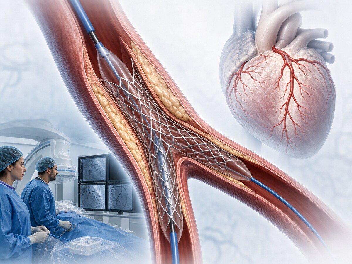
A bifurcation is a narrowing where a coronary artery divides into a main branch and a side branch. The left main coronary artery (LMCA) supplies most of the heart muscle; in trial populations the disease often sits exactly at that divide. Treatment is not always two stents and not always bypass: for most bifurcations the plan starts with one stent; a second stent, imaging and a surgical option depend on anatomy and a Heart Team discussion.[1–4,11,12]
What is a coronary bifurcation?
The European Bifurcation Club defines a bifurcation lesion as a coronary narrowing adjacent to, and/or involving, the origin of a significant side branch. In plain language, the narrowing sits at or next to the fork. That definition does not mean every bifurcation is automatically more dangerous than any other narrowing.[1]
When is a side branch called “important”?
Importance is not decided by millimetre diameter alone. The EBC counts a branch you would not want to lose, or one that needs treatment. The judgement depends on the muscle it supplies, flow and the live procedural picture; a fixed size cut-off does not decide the case on its own.[1]
What does the Medina classification mean?
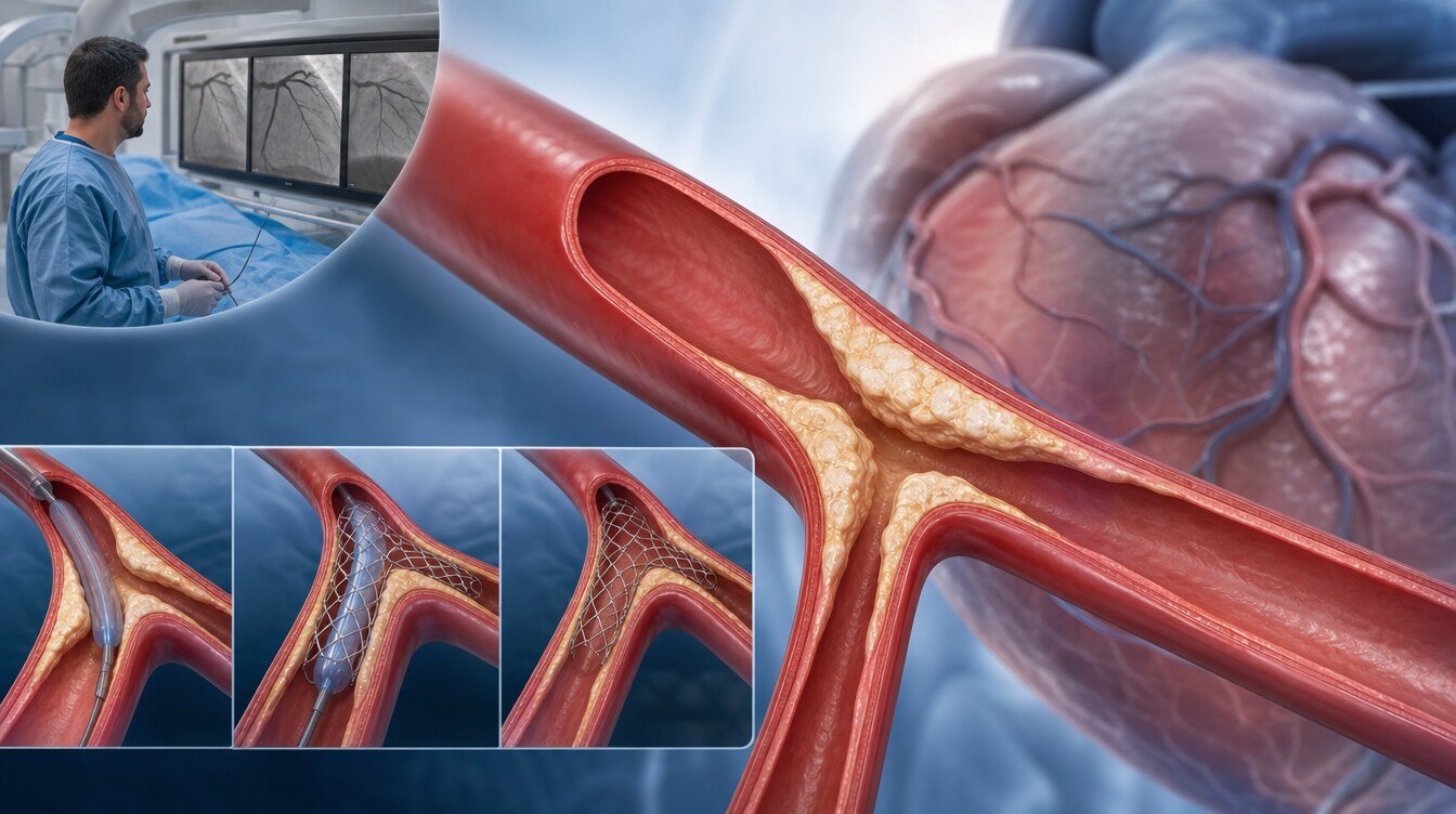
Doctors describe a bifurcation with three numbers: proximal main vessel, distal main branch and side branch. 1 means that segment has a stenosis above 50%; 0 means it does not. The order is fixed. Medina is not a severity score or a prognosis; it records which of the three parts is involved.[1]
Bif-ARC later proposed defining segment significance on imaging by a minimum lumen area below 6.0 mm² for left main segments and below 4.0 mm² elsewhere. That is not a replacement “score” for the Medina code.[2]
What is a “true” bifurcation?
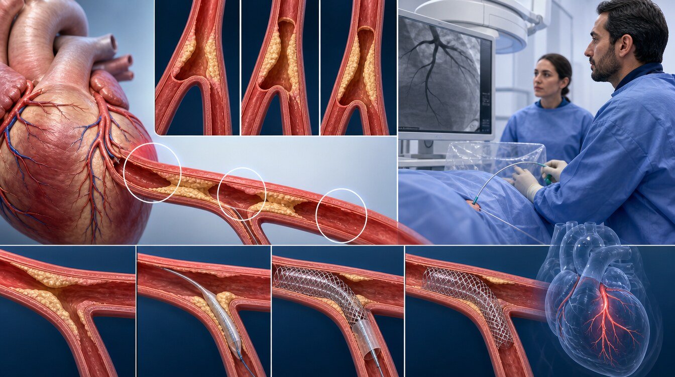
A true bifurcation means the narrowing also involves the side-branch opening: Medina 1,1,1 / 1,0,1 / 0,1,1. That does not mean two stents are required. Complexity is judged on extra criteria such as side-branch lesion length, calcium and angle.[2,5]
Why is the left main (LMCA) discussed separately?
The left main supplies most of the heart muscle. In the EXCEL angiographic core-laboratory analysis, 84.2% of assessable patients had disease involving the distal left main bifurcation versus 15.8% confined to the ostium or shaft. Those proportions describe a selected low-to-intermediate complexity trial population, not the frequency of left main disease in the general population.[4]
No randomised technique evidence specific to protected left main disease was identified in this package. The randomised left-main stent-versus-bypass and technique trials enrolled unprotected disease.[4,6,11]
Why is treatment technically demanding?
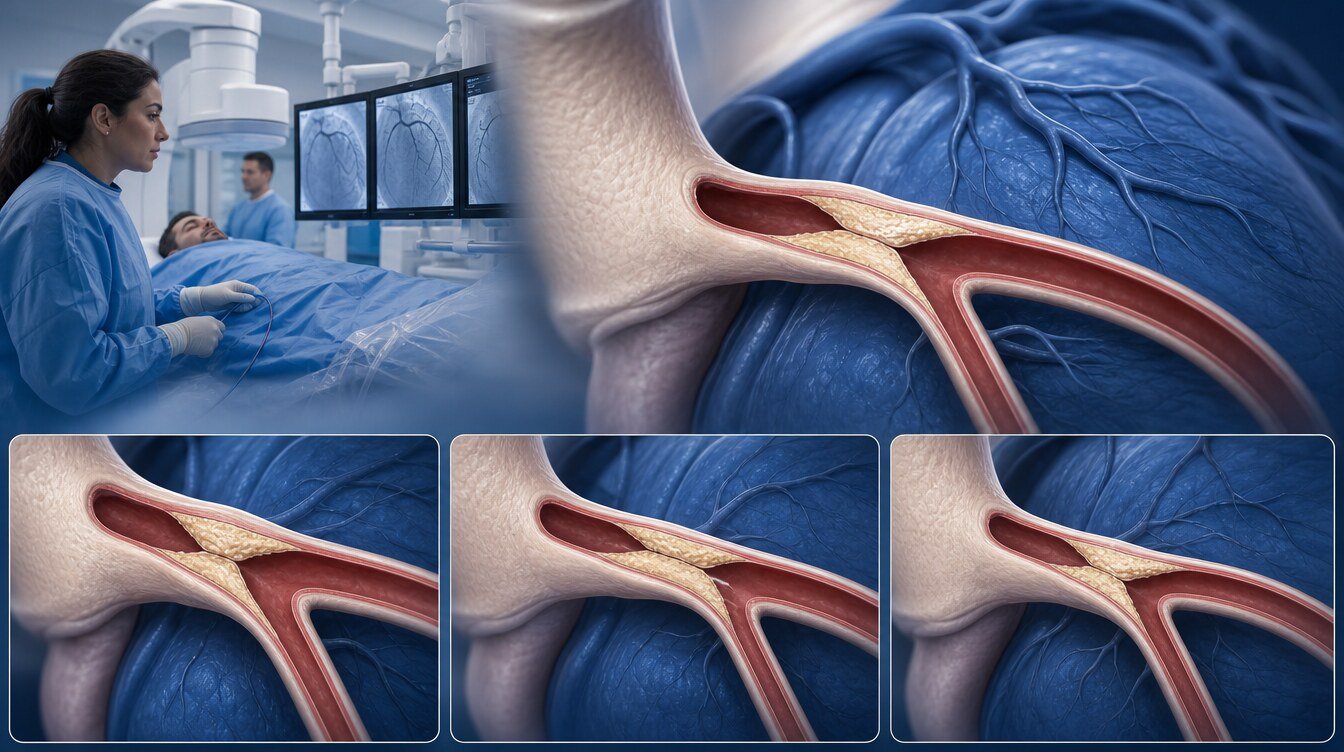
In almost every bifurcation procedure a stent sits across the take-off of a branch, so one device must serve two segments of different calibre. The EBC describes sizing the stent to the distal main vessel and then expanding the proximal segment with proximal optimisation (POT). That is a mechanical explanation, not a personal risk score or an operator checklist.[3]
One stent or two?
The EBC 18th consensus states that limiting the number of stents through a stepwise provisional strategy remains the recommended approach for the majority of true left main and non-left-main bifurcations. Across the randomised trials a second stent was needed in about 20% of patients allocated to that plan. The figure comes from selected trial populations and varies by anatomy and centre. It is not a promise that only one stent will be used.[3]
When the side branch is substantially diseased over a longer segment, a planned two-stent strategy has been linked to fewer repeat problems in that selected setting. DEFINITION II found 1-year target lesion failure of 11.4% with provisional versus 6.1% with systematic two stents in 653 patients. The DKCRUSH-X pooled analysis combines 1,573 patients already counted in the component DKCRUSH trials; 6-year target lesion failure was 18.2% with two stents versus 24.7% with provisional. Two stents are not better in general and have not been shown to reduce death.[5,6]
How are the four techniques chosen?
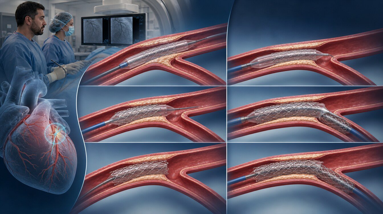
Two-stent techniques are not interchangeable “better/worse” rankings. Anatomy, operator and centre experience are reviewed together. For DK crush the EBC emphasises left main lesions treated by high-volume operators. No verified Class/LOE for DK crush, culotte or TAP was found in the retrieved guideline texts; the technical frame is consensus.[3,12]
Provisional stenting
A philosophy of main-vessel stenting, POT and a second stent only if the side branch clearly needs it — not a “one-stent guarantee”. Detail: provisional stenting.
DK crush
A planned two-stent technique. In true distal left main trials DKCRUSH-V and EBC MAIN point in different directions; no universal “best technique” claim is made. Detail: DK crush.
Culotte
Fully covers both ostia and leaves a double metal layer proximally. There is no universal ranking versus crush. Detail: culotte.
T-stenting and TAP
TAP is a modification of T-stenting used when a side-branch stent becomes necessary during a provisional procedure. There is no large randomised TAP-versus-DK crush superiority trial. Detail: T-stenting.
Why may IVUS or OCT be used during the procedure?
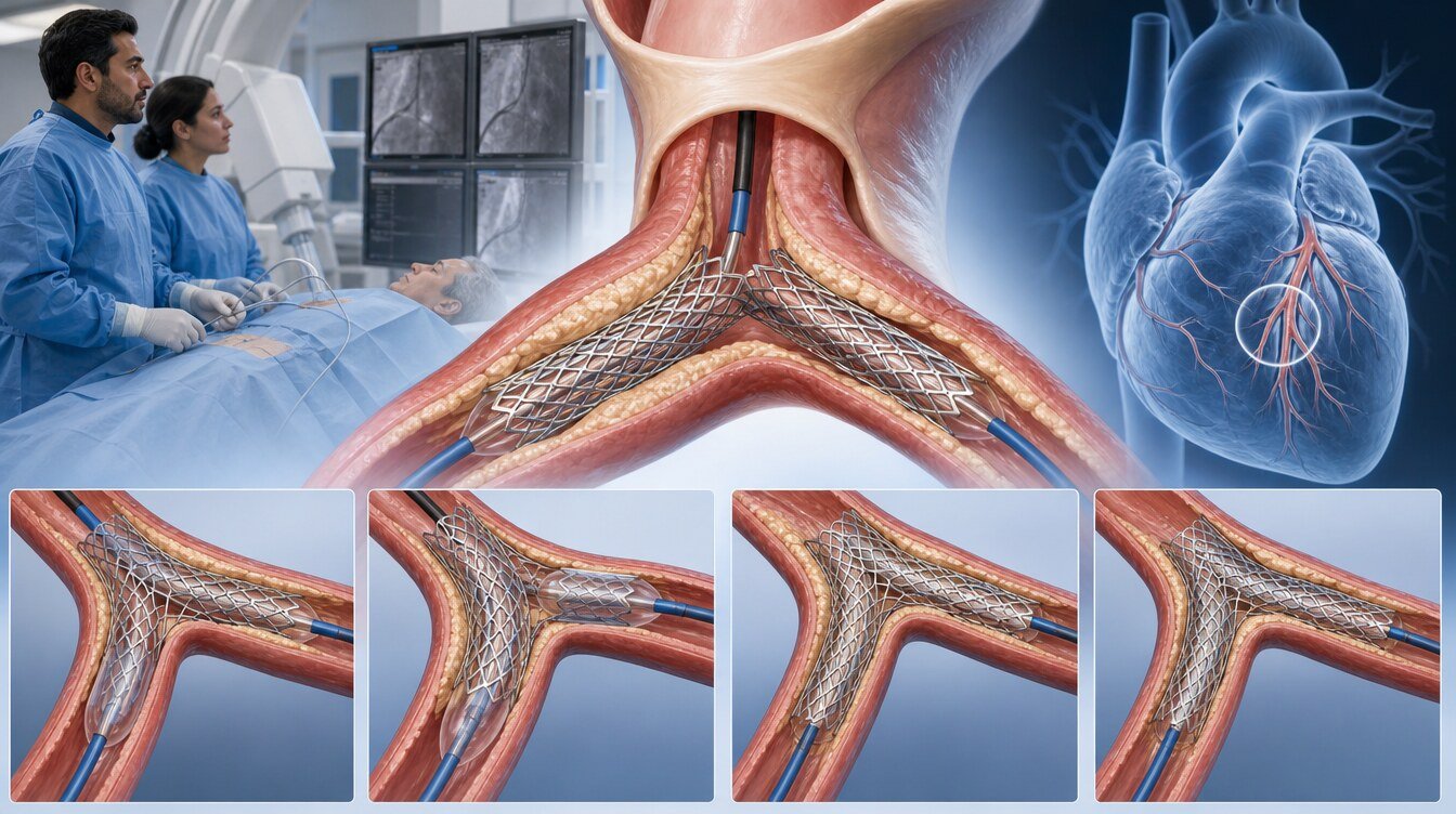
Coronary angiography gives a two-dimensional lumen view. IVUS and OCT provide cross-sections that can help assess stent apposition and plaque extent.
The 2024 ESC chronic coronary syndromes guideline recommends IVUS or OCT guidance for anatomically complex lesions, naming left main stem, true bifurcations and long lesions, at Class I Level A. The 2021 ACC/AHA/SCAI guideline grades IVUS 2a in this setting and OCT as a reasonable alternative except in ostial left main disease. That ostial left main exception is a real limitation of OCT.[7,12]
In OCTOBER, 2-year major events were 10.1% with OCT guidance versus 14.1% with angiography in complex bifurcation PCI. In the left-main substudy the difference was not statistically significant (14.4% vs 18.4%). ILUMIEN IV found a larger minimum stent area with OCT but no significant 2-year target-vessel-failure difference (7.4% vs 8.2%). Imaging can improve the stent result; it must not be said to prevent heart attack or death.[8–10]
Stent or bypass for left main disease?
This question is separate from which stenting technique to use. In an individual-patient meta-analysis of four randomised trials (4,394 patients, median SYNTAX 25), 5-year all-cause death was 11.2% with PCI and 10.2% with coronary bypass (CABG) — a non-significant difference. Spontaneous myocardial infarction (6.2% vs 2.6%) and repeat revascularisation (18.3% vs 10.7%) were more frequent after stenting. The authors add that a small mortality difference favouring CABG probably exists, most likely under 0.2% per year. The trials enrolled low-to-intermediate anatomical complexity; guidelines prefer surgery in high-complexity left main disease.[11,12]
The 2021 ACC/AHA/SCAI guideline recommends CABG to improve survival in significant left main stenosis (Class 1) and recommends choosing CABG over PCI when left main disease is accompanied by high-complexity coronary disease (Class 1). The 2024 ESC guideline treats CABG as the overall preferred revascularisation mode in low-surgical-risk patients and accepts PCI as an alternative when complexity is low (SYNTAX ≤22) and completeness can be matched. A score does not replace the discussion.[7,12]
A longer reading of the stent-versus-bypass balance is in the health-guide article Bifurcation / LMCA disease: stent or bypass?.

How is the decision made?
When the best option is unclear, guidelines recommend a Heart Team including interventional cardiology, cardiac surgery and clinical cardiology. The 2024 ESC guideline recommends calculating the SYNTAX score for anatomical complexity (Class I, B) and the STS score for early surgical risk (Class I, B). Scores support the discussion; they do not dictate treatment. Diabetes with multivessel disease has a separate guideline frame and is not merged here with left-main technique choice.[7,12]
If the lesion is heavily calcified or chronically occluded, the plan may change. Those topics have their own pages: calcified coronary lesion treatment, chronic total occlusion, stent.
How is the procedure performed?
According to the NHS, coronary angioplasty is done in a catheter laboratory rather than an operating theatre, under local anaesthetic, with the patient awake. A thin tube is passed from the wrist, arm or groin. Sedation practice varies; no fixed procedure duration is published.[13]
No official fasting interval was retrieved in this package. Follow the hospital’s instructions; a number of hours is not invented here.
The access pathway is conceptually that of coronary angiography; bifurcation or left main anatomy can make the plan more complex.
What are the risks?
The NHS lists bruising or bleeding at the puncture site as common, and arterial injury, contrast reaction, major bleeding, heart attack, stroke or death as less common but serious. The NHLBI adds arrhythmia, infection and, rarely, contrast-related kidney injury. No numerical patient-level rates are given. Risk varies with age, overall health, other conditions and whether the procedure was planned or emergency. Trial complication rates are not this patient’s risk; the procedure is not risk-free.[14,17]
Hospital stay, return to work and driving
After planned, non-emergency angioplasty the NHS states that most people can leave the same day or the next day. No retrieved official source gives a length of stay for complex left main or two-stent bifurcation PCI; same-day discharge is not promised.[15]
Official sources disagree on work and driving. The NHS advises not driving for a week after planned angioplasty and returning to work after about a week, or weeks to months after emergency angioplasty. MedlinePlus gives 2–3 days if the work is not heavy. Turkish driving rules were not retrieved in this package. Your own timing is set by your doctor; a single number is not imposed.[15,16]
Access-site care differs for wrist and groin. MedlinePlus advises keeping the site dry for 24–48 hours and limiting heavy lifting; the NHS advises avoiding heavy effort for about a week or until the wound has healed. These timings describe uncomplicated elective PCI.[15,16]
Blood-thinning medicines
The NHS states that most people take two antiplatelet medicines for up to a year after angioplasty, then usually continue low-dose aspirin long term. MedlinePlus stresses not stopping these medicines without advice, because a clot can lead to a heart attack. The 2024 ESC lists complex left main PCI and two-stent bifurcation among high-thrombotic-risk examples; that is not a personalised duration. Duration is individualised on ischaemic and bleeding risk.[7,15,16]
When should I call 112?
Call 112 and do not drive yourself for bleeding that will not stop or restarts after 10 minutes of pressure, severe chest pain that does not settle, or a cold, pale or numb limb on the puncture side. Contact the hospital or your doctor for increasing wound pain, swelling, fever, redness or discharge.[15,16]
A stent treats the narrowing, not the underlying disease. Arteries may narrow again. Medication, lifestyle change and, when offered, cardiac rehabilitation remain important.[16]
Identity-verified publications by Prof. Dr. Hakan Uçar in this field include the modified flower petal technique in Medina 0,0,1 or 0,1,0 ostial lesions (EuroIntervention, 2015; the published title is used), a jailed semi-inflated balloon series for side-branch patency, and an observational comparison of crossover versus focal ostial LAD stenting. These are single-centre or case-level; they are not evidence of superiority, a personal success rate or a treatment recommendation.[18]
Frequently asked questions
No. For most bifurcations the plan starts with one stent. In the trials about one in five patients allocated to that plan needed a second stent; the rate varies with anatomy.[3]
No. The three numbers record which segments are narrowed. It is not a severity score or a prognosis.[1]
No official fasting interval was retrieved in this package. Follow the instructions from the hospital performing your procedure.
For planned, uncomplicated angioplasty the NHS describes the same day or the next day. That is not a promise after complex left main or two-stent PCI.[15]
Call 112 for bleeding that will not stop, severe chest pain that does not settle, or a cold, pale or numb limb on the puncture side.[15]
Assessment and decision
Bifurcation and left main interventions weigh fork anatomy, the clinical importance of the side branch, imaging and, when relevant, a surgical option. A provisional approach is the starting plan for most patients; DK crush, culotte and T/TAP are not interchangeable. The decision is settled with a Heart Team and your priorities.[3,7,11]
Literature
References
Louvard Y, et al. Classification of coronary artery bifurcation lesions and treatments: time for a consensus! Catheter Cardiovasc Interv. 2008. DOI and PMID were not verified in this package.
Albiero R, et al. EBC 16th consensus, part I. EuroIntervention. 2022. DOI: 10.4244/eij-d-22-00165. PMID: 35570748.
Burzotta F, et al. EBC 18th consensus. EuroIntervention. 2024. DOI: 10.4244/eij-d-24-00160.
Gershlick AH, et al. EXCEL lesion-site analysis. J Am Coll Cardiol. 2018. PMID: 29976358. Same EXCEL cohort.
Zhang J, et al. DEFINITION II. Eur Heart J. 2020. DOI: 10.1093/eurheartj/ehaa543. PMID: 32588060. NCT02284750.
Chen SL, et al. DKCRUSH-X pooled IPD. AsiaIntervention. 2025. DOI: 10.4244/aij-d-25-00021. Overlaps component DKCRUSH trials.
Vrints C, et al. 2024 ESC Guidelines for the management of chronic coronary syndromes. Eur Heart J. 2024. Guideline text. DOI and PMID were not verified in this package.
Holm NR, et al. OCTOBER. N Engl J Med. 2023. DOI: 10.1056/NEJMoa2307770. PMID: 37634149.
Holck EN, et al. OCTOBER left main substudy. EuroIntervention. 2026. DOI: 10.4244/eij-d-25-01337. Subset of OCTOBER.
Ali ZA, et al. ILUMIEN IV. N Engl J Med. 2023. DOI: 10.1056/NEJMoa2305861. PMID: 37634188.
Sabatine MS, et al. PCI versus CABG in left main disease: IPD meta-analysis. Lancet. 2021. PMID: 34793745. Pools SYNTAX, PRECOMBAT, NOBLE and EXCEL.
Writing Committee Members. 2021 ACC/AHA/SCAI Coronary Artery Revascularization Guideline. J Am Coll Cardiol. 2021. Guideline text. DOI and PMID were not verified in this package.
NHS. Coronary angioplasty — How it is performed. Official patient page. Accessed 27 August 2026.
NHS. Coronary angioplasty — Risks. Official patient page. Accessed 27 August 2026.
NHS. Coronary angioplasty — Recovery. Official patient page. Accessed 27 August 2026.
MedlinePlus. Angioplasty and stent - heart - discharge. Official patient page. Accessed 27 August 2026.
NHLBI. Stents: After You Get a Stent. Official patient page. Accessed 27 August 2026.
Cayli M, Elbasan Z, Gur M, Seker T, Ucar H, et al. Modified flower petal technique in the treatment of Medina type 0,0,1 or 0,1,0 lesions. EuroIntervention. 2015. DOI: 10.4244/EIJV11I7A154. Single-centre technical/observational study; not evidence of superiority. The published title is used.
This content is for general information only and does not replace personalized medical advice. In emergencies call local emergency services.
Continue